

Standing Out in the Crowd: Signal-to-Background in Molecular Imaging
James R. Johnson A and David Piwnica-Worms A BA The BRIGHT Institute, Washington University School of Medicine, and Molecular Imaging Center, Mallinckrodt Institute of Radiology, Washington University School of Medicine, and Departments of Cell Biology & Physiology and Developmental Biology, Washington University School of Medicine, St. Louis, MO 63110, USA.
B Corresponding author. Email: piwnica-wormsd@mir.wustl.edu
James R. Johnson is currently a post-doctoral research scholar at the Washington University School of Medicine in Saint Louis, Missouri, USA, under the mentorship of Dr. David Piwnica-Worms. He earned his Ph.D. in 2008 from The University of Notre Dame under the advisory of Dr. Bradley Smith for his thesis ‘Targeted Delivery of Molecular Cargo and Fluorescent Bioimaging Agents’. His current research focuses on synthesis, development and validation of activatable fluorescent probes for in vitro and in vivo optical imaging applications. |

David Piwnica-Worms, M.D., Ph.D., is Professor of Radiology, Cell Biology & Physiology, and Developmental Biology, Director of the BRIGHT Institute and Director of the Molecular Imaging Center at Washington University School of Medicine, St. Louis, Missouri, USA. He earned his bachelor's degree from Stanford University, and graduated from the Duke University Medical Scientist Training Program. He continued his training at the Brigham & Women's Hospital in Diagnostic Radiology and began his first faculty appointments at Harvard Medical School. After rising to Associate Professor, Dr. Piwnica-Worms was recruited to Washington University School of Medicine and the Mallinckrodt Institute of Radiology. Dr. Piwnica-Worms has been a pioneer of the field of molecular imaging. His research interests encompass applications of molecular cell biology, high throughput screens, and chemistry to molecular imaging in vivo with specific focus on development of genetically-encoded reporter systems for imaging gene expression in vivo, imaging signal transduction and protein-protein interactions in whole cells and animal models, and biochemical analysis of the function and regulation of the multidrug resistance P-glycoprotein family of transporters. He also pursues translational research directed toward imaging in vivo using activatable optical probes, as well as technetium-99m and gallium-68 labeled radiopharmaceuticals in nuclear medicine applications. He is a founding member and former president of the Society for Molecular Imaging, and recipient of the Society for Molecular Imaging Lifetime Achievement Award as well as the Distinguished Alumnus Award, Duke University Medical School. |
Australian Journal of Chemistry 64(5) 590-592 https://doi.org/10.1071/CH11126
Published: 30 May 2011
Molecular imaging is a rapidly expanding interdisciplinary field historically focussed on the concept of non-invasively quantifying and visualizing molecular and cellular processes, rather than anatomy, as they occur in biological systems ranging in scale from the subcellular to whole organism through use of external imaging devices.[1] More recent advances have come to include minimally-invasive strategies for imaging molecular activities in situ (e.g. endoscopic or intravascular fluorescence imaging) or imaging open surgical fields, often following the administration of an appropriate targeted contrast agent.[2] Molecular imaging often utilizes the distinct chemical nature of unique injectable probes (contrast agents) to interrogate specific signalling events and receptors on and within cells, tissues, and animals. These probes or reporters can be specifically tailored to obtain quantitative and qualitative information about specific pharmacological targets and biochemical events in biological systems. The conceptual framework for molecular imaging originated in nuclear medicine with radiotracer agents in the 1970s and 1980s as the need to better understand metabolic pathways (glucose metabolism) and drug pharmacokinetics in vivo drove the development of molecular-targeted tracer compounds and imaging techniques.[3] Merging the conceptual framework of molecular-cell biology with genetically-encoded constructs or reporters and the analysis of signalling pathways and protein function in vivo with non-invasive external imaging devices spawned a leap in the mid-late 1990s towards the full scope of modern molecular imaging as now envisioned and practiced.[1]
The non-invasive character of molecular imaging is highly advantageous as it minimizes perturbation of the biological system under interrogation, allowing for more complete and accurate observations to be made in the proper context of a live subject.[1,4–7] Importantly, many gene expression profiles, signal transduction pathways, and protein–protein interactions arise from cell-mediated signalling in a tissue-restricted manner that we now understand are context-specific. For example, the functional consequences of a given gene expressed during development can be quite different when the same gene is expressed in the adult, as seen with embryonic genes that are re-expressed in cancer cells.[8] Indeed, cell autonomous genetic changes within an incipient cancer cell combined with alterations in the stromal microenvironment contribute to neoplastic progression.[9] The importance of the microenvironment in neoplastic progression is underscored by studies demonstrating that fibroblasts isolated from a tumour stimulate growth of preneoplastic and neoplastic cells in xenograft models. Thus, as this example in cancer illustrates, there is increasing need for studies of the genetic and molecular basis of disease to migrate to the whole organism to correctly capture relevant molecular mechanisms in the proper context. Molecular imaging provides one such platform for non-invasive analysis of biology in vivo. This new set of molecular probes, detection technologies and imaging strategies, collectively termed molecular imaging, now provides researchers and clinicians alike, new opportunities to visualize gene expression, biochemical reactions, signal transduction, protein–protein interactions, regulatory pathways, cell trafficking and drug action non-invasively and repetitively in their normal physiological context within living organisms in vivo. This has led to improvements in basic investigations of biomedicine in model organisms and it is hoped that this will lead to improved diagnosis and treatment of a wide variety of diseases such as cancer, inflammation, neurodegenerative disorders, and cardiovascular disease. Additionally, since the biological system of interest is left unperturbed, longitudinal studies using the same individual test subject are enabled, thereby decreasing biological variation, enhancing statistical robustness, providing new opportunities for unique protocols, and reducing the consumption of materials and laboratory animals.
Within molecular imaging are families of non-invasive imaging modalities, for example, optical fluorescence and bioluminescence imaging, magnetic resonance imaging (MRI), positron emission tomography, and single photon emission computed tomography. Molecular imaging has gained collectively from advances in molecular and cell biology, development of new molecular probes which serve as sources of imaging contrast, and in advances in imaging instrumentation. Imperative to the efficacy of molecular imaging techniques is the facile detection of these probes by their respective imaging instrumentation. Each modality has its own strengths and weaknesses for analyzing subjects of interest.[1,10] Regardless of modality, one of the largest areas remaining for technical progress in molecular imaging is overcoming limitations imposed by low signal-to-background ratios.
Signal starts with molecular probes, which provide image contrast and consist of three basic elements: a targeting moiety, a linker, and a reporter. Most labelled probes, and in particular, radiolabelled probes, are constitutive (always on), but some probes (e.g. optical, MRI) are amenable to inducible or activatable strategies. These activatable or ‘smart’ probes will generally possess similar scaffolds as standard probes, but will incorporate structural characteristics that make them active only in the presence of the target or signalling event of interest. Thus, signal from non-specific binding and uncleared non-target areas becomes less significant, allowing for greater signal-to-background ratios, enhanced temporal resolution, and multi-time point analysis with a single administration of probe.
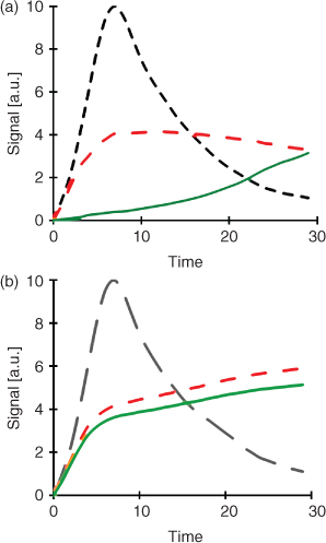
More than simply generating high target signals, molecular imaging demands high signal-to-background ratios. It is the signal-to-background ratio that determines the conspicuity of the target during whole body imaging. Thus, a great deal of effort is actually directed to ensuring rapid clearance of probes from non-target tissues and cells during a molecular imaging study. The requirements for clearance of off-target signal can vary depending on whether probes are constitutive or activatable. For constitutive agents, the overall signal-to-background ratios are critically dependent on both high targeting as well as rapid clearance from non-target tissues. This is especially true for short lived isotopes which place high demands on rapid clearance within the time frame that the target remains imagable (Fig. 1a). However, activatable or quenched optical probes hold an advantage over constitutive agents in that the requirements for non-target binding or accumulation can be relaxed somewhat, since the probe should not be visible, despite its presence at non-target sites (Fig. 1b). Thus, for an activatable probe, high target binding and activation are the primary drivers, as long as general metabolism and toxicity do not interfere with the generation of high signal-to-background ratios for imaging. Clearance, in general, is quite complex and relates to the structural details, size, surface charge, protein binding characteristics, rates of metabolism versus stability, and route of excretion of the probe. And, of course, these clearance characteristics cannot interfere with the targeting properties of the compound. Regardless of modality or probe emission properties, ideal reporters should have no toxicity, quick, and selective localization to their targets, minimal interaction with non-targets, rapid systemic clearance and, if applicable, only activate in the presence of the intended target. While these are general characteristics that should be considered for designing and synthesizing molecular imaging agents, each modality has its own unique constraints that require attention along the pathway to fulfilling these requirements. Thus, the formidable challenges for chemists in molecular imaging are to both optimize the targeting properties as well as minimize off-target binding capacity of candidate probes, whether the sources of these non-targeting characteristics reside in serum proteins, hydrophobic compartments (cell membranes, fatty tissue), off-target receptors, or metabolic pathways.
|
The field of molecular imaging is continuously expanding our ability to explore and interrogate the nature of biological systems without disrupting their integral context. Refinement of probe targeting, activation, and pharmacokinetics will continue to improve signal-to-background ratios, furthering what can be accurately detected and analyzed. Continued combination and integration of separate imaging methods into multimodal techniques and systems will enable more complete understanding of the true nature of living systems on both a micro and macro scale, all within the context of the intact biological system. Additionally, molecular imaging is an inherently interdisciplinary field and it will prosper best through increased collaboration and outreach to different fields that can add new perspectives for translation from basic science to clinical application. Regardless of modality or combination thereof, one of the central objectives to expanding the application of molecular imaging will be use of creative chemistry in overcoming limits imposed by suboptimal signal-to-background ratios.
Acknowledgements
This project was supported in part by NIH grants F32 EY20051–01 (J.R.J.), R01 EY019587, and P50 CA94056 (D.P.-W.).
References
[1] S. Gross, D. Piwnica-Worms, Cancer Cell 2005, 1, 5.[2] M. A. Whitney, J. L. Crisp, L. T. Nguyen, B. Friedman, L. A. Gross, P. Steinbach, R. Y. Tsien, Q. T. Nguyen, Nature Biotech. 2011, in press. 10.1038/NBT.1764
[3] D. Bailey, D. Townsend, P. Valk, M. Maisey, Positron Emission Tomography: Principles and Practice 2003, p. 884 (Springer-Verlag: London).
[4] R. Singer, D. Lawrence, B. Ovryn, J. Condeelis, J. Biomed. Opt. 2005, 10, 051406.
| Crossref | GoogleScholarGoogle Scholar | 16292943PubMed |
[5] V. Villalobos, S. Naik, D. Piwnica-Worms, Annu. Rev. Biomed. Eng. 2007, 9, 321.
| Crossref | GoogleScholarGoogle Scholar | 1:CAS:528:DC%2BD2sXhtVehtLbE&md5=e68424e10883e15f3bfdcebbcd58878bCAS | 17461729PubMed |
[6] R. Weissleder, M. J. Pittet, Nature 2008, 452, 580.
| Crossref | GoogleScholarGoogle Scholar | 1:CAS:528:DC%2BD1cXktFCgt7Y%3D&md5=996e84a3e5de282fdb5e6573f0c1bec4CAS | 18385732PubMed |
[7] R. Dothager, K. Flentie, B. Moss, M. Pan, A. Kesarwala, D. Piwnica-Worms, Curr. Opin. Biotechnol. 2009, 20, 45.
| Crossref | GoogleScholarGoogle Scholar | 1:CAS:528:DC%2BD1MXlslWgsrg%3D&md5=4cafefaec988832648f25913584a1cbeCAS | 19233638PubMed |
[8] M. Monk, C. Holding, Oncogene 2001, 20, 8085.
| Crossref | GoogleScholarGoogle Scholar | 1:CAS:528:DC%2BD3MXptlKktb8%3D&md5=8fa83d90f834e5d39baf11be5de9655cCAS | 11781821PubMed |
[9] M. Bissell, D. Radisky, Nat. Rev. Cancer 2001, 1, 46.
| Crossref | GoogleScholarGoogle Scholar | 1:STN:280:DC%2BD387mvVOhtw%3D%3D&md5=3506d83d9793ed5608469d5a4d5a4355CAS | 11900251PubMed |
[10] J. Condeelis, R. Weissleder, Cold Spring Harb. Perspect. Biol. 2010, 2, a003848.
| Crossref | GoogleScholarGoogle Scholar | 1:CAS:528:DC%2BC3MXhsFKqtLg%3D&md5=b8cf308fc642edb588bf05f636d4ae7fCAS | 20861158PubMed |