

Methylmercury exposure and health effects in humans
Anna L. Choi A C and Philippe Grandjean A BA Department of Environmental Health, Harvard School of Public Health, Boston, MA 02215, USA.
B Department of Environmental Medicine, University of Southern Denmark, Odense, Denmark.
C Corresponding author. Email: achoi@hsph.harvard.edu
Environmental Chemistry 5(2) 112-120 https://doi.org/10.1071/EN08014
Submitted: 12 February 2008 Accepted: 20 March 2008 Published: 17 April 2008
Environmental Context. Dietary intake of fish and other seafood products is the dominant source of human exposure to methylmercury, a toxicant that can have serious adverse effects on the developing nervous system and may promote heart diseases. The existing evidence of human toxicity should inspire prudent choices in maintaining fish intakes to secure an ample supply of essential nutrients, while at the same time choosing species that are low in mercury concentrations. The combination of essential nutrients and avoidance of this toxic contaminant will benefit brain development and human health in general. In addition, current contamination levels suggest that intensified efforts are needed to reduce and eliminate mercury release to the environment.
Abstract. Methylmercury (MeHg) is a worldwide contaminant found in seafood and freshwater fish, which constitute the dominant source of human exposure to this substance. The developing human brain is particularly susceptible to injury caused by MeHg, which easily passes the placental barrier. Epidemiological studies in fishing populations have found subtle though lasting adverse effects on brain functions of children who were exposed prenatally to MeHg from seafood diets. This contaminant also seems capable of promoting the development of heart disease. Fish and seafood also contain important nutrients, such as omega-3 fatty acids that may provide beneficial effects, thereby possibly counteracting or obscuring the adverse effects of MeHg. This article reviews the existing evidence on MeHg developmental neurotoxicity and the emerging evidence that MeHg may promote the development of heart diseases. MeHg risks may have been underestimated in the past, in part because of the confounding effects of nutrients from seafood and fish. Improved control of mercury release to the environment is indicated. In addition, regulatory agencies should provide better guidance to consumers in maintaining a balanced diet that includes seafood as low as possible in mercury.
Additional keywords: diet, environmental exposure, fish, methylmercury compounds, neuropsychological tests, seafood.
Introduction
Mercury (Hg) is a widespread contaminant found in the environment in both metallic form and in various inorganic and organic complexes.[1] The natural global biogeochemical cycling of Hg includes a conversion of Hg from inorganic into an organic form (methylmercury, MeHg) primarily in microorganisms in the aquatic environment; they are consumed by fish, and MeHg is biomagnified in the aquatic food chains so that the highest concentrations occur in large and long-lived predatory fish and marine mammals at the top trophic levels.[1]
Increased exposures to inorganic Hg may occur in connection with industrial processes, such as mercury mining, mercury catalysts at chloralkali plants, fluorescent lamp factories, and artesanal and small-scale gold mining operations.[2,3] Absorption of Hg vapor from dental amalgams is the most important source in the general population. Current concerns related to human mercury exposures particularly emphasise dietary intakes of fish and other seafood products as the dominant source of human exposure to MeHg.
MeHg poisoning outbreaks in Minamata, Japan and Iraq occurred between 1950 and 1975. Poisoning in Minamata occurred because of long-term exposure through the ingestion of fish contaminated by MeHg.[4] In Iraq, exposure was a result of the consumption of homemade bread that was made with grain treated with MeHg as fungicide.[5] Neurologic signs caused by MeHg poisoning include paresthesia, ataxia, sensory disturbances, tremors, impairment of hearing, and walking difficulties. The effects in offspring who were exposed to MeHg in utero were more serious, as the severe and widespread damage that may occur to the brain when exposed to MeHg during development were clearly demonstrated in the poisoning outbreaks. The Minamata infants were born with serious neurological damage, even if their exposed mothers were virtually unaffected.[4,6] Recent epidemiological studies have found more subtle adverse effects on brain functions at lower levels of MeHg.[7,8] A recent case-control study found that an increased blood Hg concentration was associated with attention-deficit hyperactivity disorder.[9] Although inconclusive, recent studies have suggested that MeHg may promote or predispose to the development of heart disease.[10,11] Increased MeHg exposure was associated with adverse cardiovascular effects in adults[12–14] and children.[15]
This paper reviews the human MeHg toxicology and the effects of MeHg exposure on neurobehavioural and cardiovascular outcomes, as based on human evidence from epidemiologic data. The paper also discusses issues in regard to exposure assessment and imprecision, and the relationship between the benefits from nutrients in fish and the risks associated with MeHg toxicity. These considerations are important for a proper evaluation of the human evidence and for resolving the important challenges in public health.
Major prospective cohort studies
Major prospective cohort studies of MeHg-exposed children have been conducted in New Zealand, the Faroe Islands, and the Seychelles. Table 1 outlines the major differences between these three prospective studies. Several smaller cohorts and cross-sectional studies have been published and support the notion of developmental neurotoxicity associated with MeHg in contaminated fish and seafood products,[1] but the main evidence comes from the three largest cohorts.

|
New Zealand
A cohort of 11 000 mothers, who gave birth to children in 1978, was initially screened.[16] Hair Hg concentrations were determined for 1000 mothers, who had consumed 3 fish meals per week during pregnancy. The high exposure group consisted of 73 mothers who had a hair Hg level above 6 μg g–1. At the first follow-up at age 4 years, high-exposure children, and reference children with lower exposure, were matched for potential confounders (i.e. mother’s ethnic group, age, child’s birthplace, and birth date). The high-exposure group showed significantly lower scores on childhood mental and motor development. A follow-up of the original cohort at age 6 years noted a three point decrement in intelligence quotient (IQ) and changes in affect in children born to women with Hg concentrations in hair greater than 6 μg g–1.[17]
Faroe Islands
The Faroe Islands are located in the North Atlantic between Norway, Shetland, and Iceland. Excess exposure to MeHg was mainly due to the traditional habit of eating meat from the pilot whale in this fishing community. Ingestion of whale blubber caused exposure to lipophilic contaminants, notably polychlorinated biphenyls (PCBs). Fish intake varied but showed a positive association with whale intake. The first birth cohort consisted of 1022 children born during a 21-month period in 1986–1987.[7] Prenatal MeHg exposure was determined from Hg concentrations in cord blood and maternal hair, both spanned a range of ~1000-fold. A total of 917 eligible children (90.3%) participated in the detailed examination at school age (7 years). At the 7-year and 14-year follow-up, decrements in attention, language, verbal memory, and to a lesser extent, in motor speed and visuospatial function were associated with prenatal MeHg exposure (controlled for age, sex, and confounders). Delayed latencies of the brainstem auditory evoked potentials, and decreased heart rate variability were also associated with mercury exposure.[7,8,18] Another prospective study (cohort 2) of 182 singleton term births were examined by the Neurological Optimality Score (NOS) at age 2 weeks. The NOS showed significant decreases at higher cord-blood Hg concentrations.[19]
Seychelles
A pilot cohort study and a main study, each with ~800 mother–child pairs, were conducted in the Seychelles, an archipelago in the Indian Ocean.[20] A hair sample was obtained from the mother six months after parturition. The hair segment that represented the pregnancy period was identified from the assumption that hair grows 1.1 cm per month. A subset of 217 children from the pilot cohort was evaluated at 66 months.[21] Maternal hair Hg was negatively associated with auditory comprehension, which remained significant after outliers were excluded. The main study included evaluation of the children at 6.5, 19, 29, and 66 months of age. No clear evidence of prenatal Hg neurotoxicity was found after adjustment for postnatal exposures.[21] Recent results show decreases in fine motor function associated with higher fetal exposure levels (≥10 μg g–1 maternal hair) at 9 years of age, from which the investigators suggest that delayed adverse effects might become apparent at exposure above 10–12 ppm as the children mature.[22]
MeHg exposure
Source of exposure
Fish and seafood provide an important pathway for human exposure to MeHg in freshwater and marine food chains, and MeHg contamination is the main reason for fishing advisories in the United States.[23] Fish, however, also contain essential nutrients that may provide beneficial effects to brain development and may help prevent cardiovascular disease, thereby counteracting or obscuring the adverse effects of MeHg.[24] All fish, however, do not contain similar proportions of Hg contamination and nutrients, as MeHg bioaccumulates through multiple levels of the aquatic food web. Total Hg concentrations vary widely across fish and shellfish, and mean values differ by as much as 100-fold.[25] Farmed fish is not free of MeHg and the concentrations depend on the presence of MeHg in the feed.[26] In assessing differences between wild and farmed fish, the European Food Safety Authority concluded that there is no general difference in safety between the two kinds of fish.[27] Still, options exist to produce farmed fish lower in MeHg by controlling the feed. Other possible sources of MeHg in the human diet include rice cultivated in Hg-contaminated areas,[28] organ meats of terrestrial animals,[29] and chicken and pork, probably as a result of using fish meal as livestock feed.[30]
Profiles of exposure
In fish-eating populations, high levels of Hg exposure have been documented. The median Hg concentration of maternal hair in the Faroe Islands was 4.5 μg g–1, with 27% above 10 μg g–1,[31] and in the Seychelles an average of 5.8 μg g–1.[32] Populations in the Amazon communities who rely on freshwater fish have median hair Hg levels range from 5 to 15 μg g–1.[33–37] In contrast, mean hair Hg levels in populations with minimal fish consumption range from 0.1 to 1.0 μg g–1, and mean blood Hg levels similarly range from 1.0 to 5.0 μg L–1.[38–41] The recent results in a national health study in the United States found that geometric mean hair mercury was 0.20 μg g–1 in women (among frequent fish consumers, the geometric mean hair mercury level was three-fold higher, 0.38 v. 0.11 μg g–1 in non-consumers) and in children 0.12 μg g–1 (the geometric mean hair mercury levels were two-fold higher, 0.16 v. 0.08 μg g–1 in non-consumers).[42] Likewise, the geometric mean blood Hg concentration was 0.34 µg L–1 in children and 1.02 µg L–1 in women; the geometric mean was almost four-fold higher among women who ate three or more servings of fish in the past 30 days compared with non-consumers (1.94 v. 0.51 µg L–1).[43] These results also showed that ~5–10% of the women of childbearing age have hair levels that exceed 1.0 μg g–1 and blood levels above 5 μg L–1. A higher percentage of excess exposures is found in Japan where more fish is consumed, and 73.3% of women have hair Hg levels above 1.0 μg g–1, and 1.7% above 5 μg g–1.[44]
The exposure depends not only on the frequency of fish intake and the size of each meal, but also on the species consumed. High concentrations of MeHg can be found in predatory fish and marine mammals in the top trophic levels, as MeHg biomagnifies in the aquatic food chains. Shark, tuna, swordfish, and tilefish are among the predatory fish with high MeHg concentrations.[45] Results of recent studies found elevated blood and hair Hg levels in Chinese adults and children.[46,47] Shark fin soup, a Chinese delicacy, has been found to be a major dietary source of MeHg exposure in this population.[46]
Biomarkers of exposure
In prospective studies, samples for mercury analysis have included maternal hair, cord blood, and cord tissue. Maternal dietary questionnaires have also been used to obtain information on the frequency of fish and seafood consumption. Blood gives an estimate of most recent exposure, with the half-life of MeHg in blood being 50–70 days, while hair may provide a calendar of Hg exposure.[32] Although hair growth rates are known to vary, a 9-cm hair sample obtained at parturition or shortly thereafter would be thought to represent the average Hg exposure during the whole pregnancy.[48] Hair Hg is predominantly MeHg (80–98% of hair total Hg). Generally, hair is 250–300 times more concentrated in Hg than in blood,[49] although cord blood contains a relative increase, thus making the difference from hair only ~180-fold.[31]
Because of the ease of sampling, storage, and transport, scalp hair is the most frequently used sample for MeHg exposure assessment.[49] However, hair Hg concentration is subject to variability such as hair type, hair color, external contamination and leaching as a result of permanent hair treatment.[50–52] These factors might well account for the greater overall imprecision of this biomarker. The blood concentration is often considered a more appropriate indicator of the absorbed dose and the amount systemically available, but this biomarker may also be subject to possible variation. The dry-weight-based mercury concentration in the cord tissue seems to be a more precise parameter than the level expressed on a wet-weight basis.[53] In assessing exposure biomarker imprecision and providing proper adjustment for its consequences, Grandjean and Budtz-Jørgensen[54] documented that cord blood is the best available indicator of prenatal MeHg exposure. However, the results also suggested that even the best exposure biomarker may be much more imprecise than suggested by laboratory quality data.
Studies of the cardiovascular effect of Hg have used Hg levels in toenails and fingernails as biomarkers of Hg exposure,[11,55] although the extent that these reflect organic or inorganic Hg exposure is unclear. The fact that dentists have increased toenail Hg concentrations[11] would suggest that this biomarker reflects not only MeHg exposure. Urinary Hg excretion levels have been used in some studies,[56,57] but the concentrations mainly reflect inorganic Hg and is not considered a useful biomarker of MeHg exposure.[57]
Health effects
Neurodevelopmental outcomes
The validity of outcome variables depends on their sensitivity to the exposure under study and the associated specificity (i.e. lack of sensitivity to the influence of other factors, including confounders). The choice of effect parameters must at the same time be feasible and appropriate for the age of the children, and for the setting of the study. Tests that depend only minimally on cooperation of the subject have the advantage of being less likely to be affected by motivation. The more advanced neuropsychological tests are only possible when a child has reached school age. Such tests, however, may be of uncertain validity if they have not previously been applied in the same culture. In addition, many tests require special skills of the examiner. All of these issues need to be considered when evaluating the study findings.[48]
Most studies employed a battery of neurobehavioural tests, some of which appeared to be more sensitive to Hg neurotoxicity than others. Simple comparison of regression coefficients may provide suggestions for the most sensitive parameter, at least within the confines of a particular study. A better sensitivity may be due to superior psychometric properties, not necessarily greater susceptibility to MeHg. To facilitate such comparison, the regression coefficient may be expressed as a proportion of the standard deviation of the test result, or as a delay in mental development calculated from the regression coefficient for age.[58]
Benchmark dose levels may also be used as a basis for comparison. Thus, the most sensitive neurological, neuropsychological, and neurophysiological effect parameters all exhibit benchmark dose levels of 5–10 μg g–1 for hair.[1] Despite the great variability of the study settings and the outcome variables, a substantial degree of concordance exists and that the combined evidence is quite convincing in regard to the dose–response relationship.
Neurological tests
Neurological examination has been included in prospective studies[19] and cross-sectional studies.[59,60] The clinical tests, however, provide only suggestive evidence to link low-dose MeHg exposures to detectable abnormalities. The absence of clear, positive findings most probably reflects the lack of sensitivity of this type of examination within this range of exposures. One weakness is that the performance on a clinical test is rated by the examiner, thereby introducing a potential subjective aspect. In addition, scoring is usually a simple pass–fail or pass–questionable-fail, thereby limiting the sensitivity.
Neuropsychological tests
While likely to be more sensitive in revealing early neurotoxic changes, neuropsychological tests require that the administration is standardised, and they may show examiner-dependence. In addition, they may be sensitive to details in the test situation, such as the use of an interpreter, differences in temperature, and other aspects that may be important when a test is used for the first time in a particular culture. In New Zealand, two psychologists tested the same children with a shortened version of the test battery and documented a high level of agreement.[17] In the Faroes and several other studies, each test was administered to all children by the same examiner, thus limiting the possible impact of examiner-related differences.
Traditionally, studies in this field have included standard intelligence batteries. For example, the Wechsler Intelligence Scale for Children (WISC) and the McCarthy Scale of Children’s Abilities were included in the New Zealand,[17] as well as the Seychelles examinations.[61,62] These intelligence tests may not be the most appropriate and sensitive for MeHg toxicity, although significant results were found on WISC and McCarthy in the New Zealand study. Some WISC subtests were used under different circumstances in the cross-sectional studies administered by an interpreter, e.g. in Madeira,[63] thereby making the test results less reliable.
In studies of dietary long-chained n-3 polyunsaturated fatty acids (LCPUFA), which are transferred via the placenta or supplied in human milk, are necessary for normal brain growth and development in infancy, and may play an important role in the development of infant cognition, the Bayley MDI performance, the Fagan Test of Infant Intelligence, and the MacArthur Communicative Development Inventory were included in the assessments on infant cognitive function.[64–66] The approach to neurobehavioural assessment taken by the Faroes was to emphasise tests that reflected functional domains (e.g. attention, motor speed, verbal memory). The functions chosen were those that were most likely to be affected by developmental MeHg exposure, as judged from location of neuropathological lesions in poisoning cases and as illustrated by studies of other developmental neurotoxicants, especially lead. The Boston Naming test (language) showed a wide range of responses and appeared to be the most sensitive outcome.[48]
Neurophysiological tests
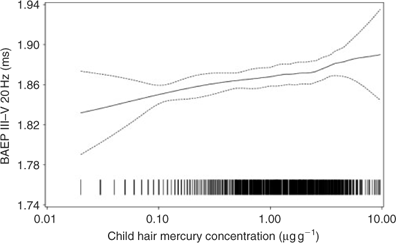
As an objective evaluation of brain dysfunction that is probably less sensitive to motivation of socioeconomic confounding, neurophysiological tests have been applied in several studies. Their applicability requires advanced instrumentation and depends on skilled examiners. An outcome that has been previously found to be sensitive to lead exposure is brainstem auditory evoked potentials (BAEP). They are recorded using surface electrodes placed on the skull while the child listens to a stimulus in one ear. The transmission of the electrical signals within the brain is then recorded as peaks that represent the acoustic nerve, an intermediate connection in the pons, and the midbrain. Patients from Minamata, Japan, with congenital MeHg poisoning exhibited delays in BAEP latencies.[67] The latency of peak III was significantly increased at higher intra-uterine exposure to mercury. Parallel associations were found in 7-year-old children in the Faores[68] and in Madeira,[69] and this observation was replicated in the Faroese cohort when examined at 14 years.[70] This study showed that delays in peak V were associated with the adolescents’ current exposure, and that early effects occurred at very low exposure levels (Fig. 1).[70] As a parameter primarily affected by postnatal exposure, peak V delays may provide unique insight in comparison with the majority of functions that are mainly sensitive to MeHg during fetal development.
|
Cardiovascular outcomes
Although the developing brain is considered the critical target organ in regard to MeHg, recent evidence has suggested that Hg from fish and seafood may promote or predispose towards the development of heart disease. This evidence is yet inconclusive, but deserves attention, because it suggests that a narrow definition of subpopulations at risk, i.e. pregnant women, might leave out other vulnerable groups.
The first studies of MeHg-associated cardiovascular disease were carried out in Finland. One important study showed that the intima-media thickness of the carotid arteries is in apparent association with the degree of mercury exposure from fish.[12] A possible mechanism could be induction of lipid peroxidation. In this regard, it is interesting to note that while essential fatty acids from fish may reduce the risk of acute coronary events, a high Hg content in fish could attenuate this beneficial effect.[71] The increased risk seems to occur at hair-mercury concentrations above 2 μg g–1, i.e. only twice the level that corresponds to the US EPA Reference Dose (a daily intake level designed to be without a significant risk of adverse effects over a lifetime). A recent study of Finnish men reported that a high mercury level was associated with an increased risk of acute coronary events and cardiovascular mortality, and that mercury may also attenuate the protective effects of fish on cardiovascular health.[13] A large multi-centre study from Europe showed an increased risk of cardiovascular disease associated with toenail mercury concentrations, and high Hg content may diminish the cardioprotective effect of fish intake.[14] A study of US health professionals did not find an association between Hg exposure and coronary heat disease.[11] However, after exclusion of dentists with high toenail Hg concentrations likely a result of amalgam exposures, the study showed an odds ratio similar to the ones seen in Finland and in the multi-centre study.[11]
Nutrients and co-contaminants
Certain essential nutrients in fish and seafood may provide beneficial effects on brain development and may protect against the development of heart disease, thereby possibly counteracting the adverse effects of MeHg. LCPUFA are essential for normal brain development and better vision,[72] and have been suggested to play a key role against cardiovascular diseases.[73,74] (Other non-fish LCPUFA sources include walnuts, flaxseed, soybeans, oil (flaxseed, canola, and soybean), and eggs.) Selenium is a trace mineral that is essential to health. Although selenium has been considered to potentially provide protection against MeHg effects, cord blood selenium concentrations in the Faroe Islands did not impact on MeHg-associated neurobehavioural deficits.[75] Iron is essential for oxygen transport and for the regulation of cell growth and differentiation,[76] and iron deficiency has adverse effects on the cognitive and psychomotor development of children.[77,78] Iodine, a trace element, is an essential component of thyroid hormones required for normal development and metabolism. Infants and children with iodine deficiency are at risk of poor mental and psychomotor development.[79] Vitamin E may interact with selenium additively because of their similar antioxidant roles. There is evidence to suggest that deficiency in Vitamin E may be associated with neurological functions in children and adults.[80,81] Although nutrients do not vary to the same extent in seafood as MeHg, it is important to control for fish intake in the study design and the statistical analysis. Failure to do so will likely result in underestimation of the MeHg toxicity.
Fish may also biomagnify persistent halogenated organic compounds, such as PCBs. The neurodevelopmental effects of PCBs share some similarities to those observed for MeHg.[82] Exposure of such toxicants, therefore, should also be considered in constructing MeHg-specific dose–response relations. For example, the Faroese are exposed to PCBs from eating whale blubber.[7] However, no important impact of PCB exposure on MeHg neurotoxicity was identified from detailed analyses of the Faroes data.[19,82,83] The relative importance of PCBs and Hg was also assessed in structural equation analyses taking into account the imprecision of the variables. The inclusion of PCB exposure attenuated the MeHg effect somewhat, but MeHg remained statistically significant, while PCB was far from that.[84] In New Zealand and Seychelles, the ocean fish consumed is unlikely to be contaminated by PCB, and the same would be the case with freshwater fish in the Amazon Basin, where MeHg neurotoxicity is also prevalent.[85]
Nutrient and MeHg exposure as predictors of developmental outcomes
Only a small number of studies have aimed at examining the effects of both nutrient and contaminant intakes at the same time as predictors of developmental outcomes. A beneficial association of the developmental score with the fish intake of the mother during pregnancy and of the infant postnatally was reported but no effect of low mercury concentrations in umbilical cord tissue (wet weight) was found.[86] However, the validity of the latter finding is uncertain because of the imprecision of the mercury exposure biomarker. In the Faroes, an adverse neonatal neurological function was associated with increased prenatal MeHg exposure, but neither n-3 fatty acids nor selenium provided any detectable beneficial or protective effect on this outcome.[19] No evidence was found that selenium provided any important protection against MeHg-associated deficits on neuropsychological tests in two separate Faroese birth cohorts.[75] In a Polish cohort, maternal fish intake during pregnancy was positively related to both maternal and cord blood mercury concentrations, which were associated with delayed psychomotor development of infants in the first year of life.[87] Three studies, in particular, showed that the effects of both nutrient and contaminant intakes were strengthened when both maternal fish intake and prenatal MeHg exposure were adjusted in modelling the same neurodevelopmental outcomes.[88–90] Thus, both the beneficial and the adverse effects of fish and seafood intake should be assessed at the same time to separate opposite impacts on the outcomes. Only then will the full impact of MeHg toxicity (and the beneficial effects of the nutrients) be apparent. As a corollary, optimising the benefits of fish and seafood consumption requires a prudent choice of species high in nutrients and as low as possible in MgHg contamination.
Conclusions
This review outlines the substantial scientific evidence on MeHg developmental neurotoxicity and the emerging evidence that MeHg may promote or predispose towards the development of heart diseases. These adverse effects are likely to occur even at exposures thought to be fairly low. The developing human brain is inherently more susceptible to injury caused by toxic agents such as MeHg than is the brain of the adult, and neurodevelopmental disorders can cause lifelong disability.[91] In terms of public health relevance, even a subtle delay in mental development may be important, especially because vast populations may be affected. The Faroes study showed that each doubling in prenatal MeHg exposure corresponded to a delay of one or two months in mental development at age 7 years,[7] which may correspond to an approximate deficit of ~1.5 IQ points, had an IQ scale been used. A similar result was seen in the New Zealand study,[17] while comparable adjusted data are not available from the Seychelles. Small shifts in a measure of the central tendency of the IQ distribution may be associated with large changes in the tails of the distribution. The lesson from lead neurotoxicity suggests that such effects are likely to be permanent and that they may even become more apparent with time.[91] Although the evidence on cardiovascular effects of MeHg is less certain, it suggests that avoidance of increased MeHg exposure would be a safe and prudent strategy for the population at large. The effect of MeHg on the human immune system has not been studied, although occupational exposure to elemental Hg has been found to alter certain immune parameters.[1]
Fish and seafood provide an important pathway for human exposures to biomagnified MeHg, but fish and marine food also contain essential nutrients that may provide beneficial health effects. In regard to existing research studies, these effects in opposite directions have served to bias the observed MeHg toxicity towards lower and less apparent levels.[89] Unfortunately, most observational studies in this field have focussed either on the risk of MeHg or on nutrient benefits. Future studies should assess both beneficial and adverse effects of fish and seafood intake, taking into account the species consumed and the consumption frequency at the same time to separate opposite impacts on the outcomes. Further, some confusion may occur because of the two different key messages. Fish and seafood provide beneficial nutrients, and the fish that are high in omega-3 fatty acids should be favoured. On the other hand, advisories against MeHg exposure should emphasise that the contamination is the greatest in larger and older fish, especially in species high in the food chain, and in those originating from contaminated waters. Fortunately, certain types of fish and seafood have a high content of beneficial nutrients but do not necessarily contain much MeHg.[45] Consumption of fish with low levels of MeHg and organic contaminants and high levels of omega-3 fatty acids constitute a ‘win win’ situation and should be encouraged regardless of the underlying nature of the omega-3–MeHg interaction.[3] Regulatory agencies, therefore, need to develop risk communication strategies for balanced messages regarding nutrients and Hg to assist the consumers in making this choice.[48]

|
Acknowledgements
This work was supported by the USA National Institute of Environmental Health Sciences (ES09797 and ES13692). The contents of this paper are solely the responsibility of the authors and do not represent the official views of the NIEHS, NIH.
[1]
[2]
[3]
D. Mergler ,
H. A. Anderson ,
L. H. M. Chan ,
K. R. Mahaffey ,
M. Murray ,
M. Sakamoto ,
A. H. Stern ,
Methylmercury exposure and health effects in humans: a worldwide concern.
Ambio 2007
, 36, 3.
| Crossref | GoogleScholarGoogle Scholar | PubMed |
[Verified 2 March 2007].
[24]
Anon, The Madison Declaration on Mercury Pollution.
Ambio 2007
, 36, 62.
| Crossref | GoogleScholarGoogle Scholar | PubMed |
[Verified 17 April 2007].
[28]
M. Horvat ,
N. Nolde ,
V. Fajon ,
V. Jereb ,
M. Logar ,
S. Lojen ,
R. Jacimovic ,
I. Falnoga ,
Q. Liya ,
J. Faqaneli ,
D. Drobne ,
Total mercury, methylmercury, and selenium in mercury polluted areas in the province Guizhou, China.
Sci. Total Environ. 2003
, 304, 231.
| Crossref | GoogleScholarGoogle Scholar | PubMed |

[29]
G. Ysart ,
P. Miller ,
M. Croasdale ,
H. Crews ,
P. Robb ,
M. Baxter ,
C. de L’Argy ,
N. Harrison ,
1997 UK Total Diet Study – dietary exposures to aluminium, arsenic, cadmium, chromium, copper, lead, mercury, nickel, selenium, tin, and zinc.
Food Addit. Contam. 2000
, 17, 775.
| Crossref | GoogleScholarGoogle Scholar | PubMed |
[30]
A. Lindberg ,
K. A. Bjornberg ,
M. Berglund ,
Exposure to methylmercury in non-fish-eating people in Sweden.
Environ. Res. 2004
, 96, 28.
| Crossref | GoogleScholarGoogle Scholar | PubMed |
[31]
P. Grandjean ,
P. Weihe ,
P. J. Jorgensen ,
T. Clarkson ,
E. Cernchiair ,
T. Videro ,
Impact of maternal seafood diet on fetal exposure to mercury, selenium and lead.
Arch. Environ. Health 1992
, 47, 185.
| PubMed |
[32]
E. Cernichiari ,
R. Brewer ,
G. J. Myers ,
D. O. Marsh ,
L. W. Lapham ,
C. Cox ,
C. F. Shamlaye ,
M. Berlin ,
P. W. Davidson ,
T. W. Clarkson ,
Monitoring methylmercury during pregnancy: maternal hair predicts fetal brain exposure.
Neurotoxicology 1995
, 16, 711.
| PubMed |
[33]
E. C. de Oliveira Santos ,
I. M. Jesus ,
E. S. Brabo ,
E. C. Loureiro ,
A. F. Mascarenhas ,
J. Weirich ,
V. M. Camara ,
D. Clearly ,
Mercury exposures in riverside Amazon communities in Para, Brazil.
Environ. Res. 2000
, 84, 100.
| Crossref | GoogleScholarGoogle Scholar | PubMed |
[34]
A. A. Boischio ,
D. S. Henshel ,
Linear regression models of methyl mercury exposure during prenatal and early postnatal life among riverside people along the Upper Madeira river, Amazon.
Environ. Res. 2000
, 83, 150.
| Crossref | GoogleScholarGoogle Scholar | PubMed |
[35]
J. Dolbec ,
D. Mergler ,
F. Larribe ,
M. Roulet ,
J. Lebel ,
M. Lucotte ,
Sequential analysis of hair mercury levels in relation to fish diet of an Amazonian population, Brazil.
Sci. Total Environ. 2001
, 271, 87.
| Crossref | GoogleScholarGoogle Scholar | PubMed |
[36]
J. Dorea ,
A. C. Barbosa ,
I. Ferrari ,
J. R. de Souza ,
Mercury in hair and in fish consumed by Riparian women of the Rio Negro, Amazon, Brazil.
Int. J. Environ. Health Res. 2003
, 13, 239.
| Crossref | GoogleScholarGoogle Scholar | PubMed |
[37]
S. Cordier ,
C. Grasmick ,
M. Paquier Passelaigue ,
L. Mandereau ,
J. P. Weber ,
M. Jouan ,
Mercury exposure in French Guiana: levels and determinants.
Arch. Environ. Health 1998
, 53, 299.
| PubMed |
[38]
A. H. Stern ,
M. Gochfeld ,
C. Weisel ,
J. Burger ,
Mercury and methylmercury expsoure in the New Jersey pregnant population.
Arch. Environ. Health 2001
, 56, 4.
| PubMed |
[39]
A. Pesch ,
M. Wilhelm ,
U. Rostek ,
N. Schmitz ,
M. Weishoff-Houben ,
U. Ranft ,
H. Idel ,
Mercury concentrations in urine, scalp hair, and saliva in Germany.
J. Expo. Anal. Environ. Epidemiol. 2002
, 12, 252.
| Crossref | GoogleScholarGoogle Scholar | PubMed |
[40]
K. A. Bjornberg ,
M. Vahter ,
K. Petersson-Grawe ,
A. Glynn ,
S. Cnattingius ,
P. P. Darnerud ,
S. Atuma ,
M. Aune ,
W. Becker ,
M. Berglund ,
Methyl mercury and inorganic mercury in Swedish pregnant women and in cord blood: influence of fish consumption.
Environ. Health Perspect. 2003
, 111, 637.
| PubMed |
[41]
L. Knobeloch ,
H. A. Anderson ,
P. Imm ,
D. Peters ,
A. Smith ,
Fish consumption, advisory awareness, and hair mercury levels among women of childbearing age.
Environ. Res. 2005
, 97, 220.
| Crossref | GoogleScholarGoogle Scholar | PubMed |
[42]
M. A. McDowell ,
C. F. Dillon ,
J. Osterloh ,
P. M. Bloder ,
E. Pellizzari ,
R. Fernando ,
R. M. De Oca ,
S. E. Schober ,
T. Sinks ,
R. L. Jones ,
K. R. Mahaffey ,
Hair mercury levels in US children and women of childbearing age: reference range data from NHANES 1999–2000.
Environ. Health Perspect. 2004
, 112, 1165.
| PubMed |
[43]
S. E. Schober ,
T. H. Sinks ,
R. L. Jones ,
P. M. Bolger ,
M. McDowell ,
J. Osterloh ,
E. S. Garrett ,
R. A. Canady ,
C. F. Dillon ,
Y. Sun ,
C. B. Joseph ,
K. A. Mahaffey ,
Blood mercury levels in US children and women of childbearing age 1999–2000.
JAMA 2003
, 289, 1667.
| Crossref | GoogleScholarGoogle Scholar | PubMed |
[44]
A. Yasutake ,
M. Matsumoto ,
M. Yamaguchi ,
N. Hachiya ,
Current hair mercury levels in Japanese for estimation of methylmercury exposure.
J. Health Sci. 2004
, 50, 120.
| Crossref | GoogleScholarGoogle Scholar |
[45]
[46]
C. M. Y. Choy ,
C. W. K. Lam ,
L. T. F. Cheung ,
C. M. Briton-Jones ,
L. P. Cheung ,
C. J. Haines ,
Infertility, blood mercury concentrations and dietary seafood consumption: a case-control study.
British J. Obstet. Gyn. 2002
, 109, 1121.
[47]
P. Ip ,
V. Wong ,
M. Ho ,
J. Lee ,
W. Wong ,
Environmental mercury exposure in children: South China’s experience.
Pediatr. Int. 2004
, 46, 715.
| Crossref | GoogleScholarGoogle Scholar | PubMed |
[48]
[49]
[50]
[51]
R. Yamamoto ,
T. Suzuki ,
Effects of artificial hair-waving on hair mercury values.
Int. Arch. Occup. Environ. Health 1978
, 42, 1.
| Crossref | GoogleScholarGoogle Scholar | PubMed |
[52]
A. Yasutake ,
M. Matsumoto ,
M. Yamaguchi ,
N. Hachiya ,
Current hair mercury levels in the Japanese: survey in five districts.
Tohoku J. Exp. Med. 2003
, 199, 161.
| Crossref | GoogleScholarGoogle Scholar | PubMed |
[53]
P. Grandjean ,
E. Budtz-Jørgensen ,
P. J. Jørgensen ,
P. Weihe ,
Total imprecision of exposure biomarkers: implications for calculating exposure limits.
Am. J. Ind. Med. 2007
, 50, 712.
| Crossref | GoogleScholarGoogle Scholar | PubMed |
[54]
P. Grandjean ,
E. Budtz-Jørgensen ,
P. J. Jørgensen ,
P. Weihe ,
Umbilical cord mercury concentration as biomarker of prenatal exposure to methylmercury.
Environ. Health Perspect. 2005
, 113, 905.
| PubMed |
[55]
E. Guallar ,
M. I. Sanz-Gallardo ,
P. van’t Veer ,
P. Bode ,
A. Aro ,
J. Gomez-Aracena ,
J. D. Kark ,
R. A. Riemersma ,
J. M. Martin-Moreno ,
F. J. Kok ,
Mercury, fish oils, and the risk of myocardial infarction.
New Engl. J. Med. 2002
, 347, 1747.
| Crossref | GoogleScholarGoogle Scholar |
[56]
T. Ohno ,
M. Sakamoto ,
T. Kurosawa ,
M. Dakeishi ,
T. Iwata ,
K. Murata ,
Total mercury levels in hair, toenail and urine among women free from occupational exposure and their relations to tubular renal functions.
Environ. Res. 2007
, 103, 191.
| Crossref | GoogleScholarGoogle Scholar | PubMed |
[57]
M. Berglund ,
B. Lind ,
K. A. Bjornberg ,
B. Palm ,
O. Einarsson ,
M. Vahter ,
Inter-individual variations of human mercury exposure biomarkers: a cross-sectional assessment.
Environ. Health 2005
, 4, 20.
| Crossref | GoogleScholarGoogle Scholar | PubMed |
[58]
P. Grandjean ,
E. Budtz-Jørgensen ,
R. F. White ,
P. J. Jørgensen ,
P. Weihe ,
F. Debes ,
N. Keiding ,
Methylmercury exposure biomarkers as indicators of neurotoxicity in children aged 7 years.
Am. J. Epidemiol. 1999
, 14, 301.
[59]
S. Cordier ,
M. Garel ,
L. Mandereau ,
H. Morcel ,
P. Doineau ,
S. Gosme-Seguret ,
D. Josse ,
R. White ,
C. Amiel-Tison ,
Neurodevelopmental investigations among methylmercury-exposed children in French Guiana.
Environ. Res. 2002
, 89, 1.
| Crossref | GoogleScholarGoogle Scholar | PubMed |
[60]
D. O. Marsh ,
M. D. Turner ,
J. C. Smith ,
V. M. H. Perez ,
P. Allen ,
N. Richdale ,
Fetal methylmercury study in a Peruvian fish-eating population.
Neurotoxicology 1995
, 16, 717.
| PubMed |
[61]
P. W. Davidson ,
J. Kost ,
G. J. Myers ,
C. Cox ,
T. W. Clarkson ,
Methylmercury and neurodevelopment: reanalysis of the Seychelles child development study outcomes at 66 months of age.
JAMA 2001
, 285, 1291.
| Crossref | GoogleScholarGoogle Scholar |
[62]
G. J. Myers ,
P. W. Davidson ,
C. Cox ,
C. F. Shamlaye ,
D. Palumbo ,
E. Cernichiari ,
J. Sloane-Reeves ,
G. E. Wilding ,
J. Kost ,
L. S. Huang ,
T. W. Clarkson ,
Prenatal methylmercury exposure from ocean fish consumption in the Seychelles child development study.
Lancet 2003
, 361, 1686.
| Crossref | GoogleScholarGoogle Scholar | PubMed |
[63]
K. Murata ,
P. Weihe ,
S. Araki ,
E. Budtz-Jørgensen ,
P. Grandjean ,
Evoked potentials in Faroese children prenatally exposed to methylmercury.
Neurotoxicol. Teratol. 1999
, 21, 471.
| Crossref | GoogleScholarGoogle Scholar | PubMed |
[64]
P. Willatts ,
J. S. Forsyth ,
The role of long-chain polyunsaturated fatty acids in infant cognitive development.
Prostaglandins Leukot. Essent. Fatty Acids 2000
, 63, 95.
| Crossref | GoogleScholarGoogle Scholar | PubMed |
[65]
E. E. Birch ,
D. G. Birch ,
D. R. Hoffman ,
R. Uauy ,
Dietary essential fatty acid supply and visual acuity development.
Invest. Ophthalmol. Vis. Sci. 1992
, 33, 3242.
| PubMed |
[66]
A. Lucas ,
R. Morley ,
T. J. Cole ,
G. Lister ,
C. Leeson-Payne ,
Breast milk and subsequent intelligence quotient in children born preterm.
Lancet 1992
, 339, 261.
| Crossref | GoogleScholarGoogle Scholar | PubMed |
[67]
R. Hamada ,
Y. Yoshida ,
A. Kuwano ,
I. Mishima ,
A. Igata ,
Auditory brainstem responses in fetal organic mercury poisoning.
Shinkei-Naika 1982
, 16, 282.
[in Japanese]
[68]
K. Murata ,
P. Weihe ,
S. Araki ,
E. Budtz-Jørgensen ,
P. Grandjean ,
Evoked potentials in Faroese children prenatally exposed to methylmercury.
Neurotoxicol. Teratol. 1999
, 21, 471.
| Crossref | GoogleScholarGoogle Scholar | PubMed |
[69]
K. Murata ,
P. Weihe ,
A. Renzoni ,
F. Debes ,
R. Vasconcelos ,
F. Zino ,
S. Araki ,
P. J. Jørgensen ,
R. F. White ,
P. Grandjean ,
Delayed evoked potentials in children exposed to methylmercury from seafood.
Neurotoxicol. Teratol. 1999
, 21, 343.
| Crossref | GoogleScholarGoogle Scholar | PubMed |
[70]
K. Murata ,
P. Weihe ,
E. Budtz-Jørgensen ,
P. J. Jørgensen ,
P. Grandjean ,
Delayed brainstem auditory evoked potential latencies in 14-year-old children exposed to methylmercury.
J. Pediatr. 2004
, 144, 177.
| Crossref | GoogleScholarGoogle Scholar | PubMed |
[71]
T. Rissanen ,
S. Voutilainen ,
K. Nyyssonen ,
T. A. Lakka ,
J. T. Salonen ,
Fish oil-derived fatty acids, docosahexaeonoic acid and docosapentaenoic acid, and the risk of acute coronary events: the Kuopio Ischaemic heart disease risk factor study.
Circulation 2000
, 102, 2677.
| PubMed |
[72]
S. M. Innis ,
Essential fatty acids in growth and development.
Prog. Lipid Res. 1991
, 30, 39.
| Crossref | GoogleScholarGoogle Scholar | PubMed |
[73]
D. Kromhout ,
F. B. Bosschieter ,
C. de Lezenne Coulander ,
The inverse relation between fish consumption and 20-year mortality from coronary heart disease.
New Engl. J. Med. 1985
, 312, 1205.
[74]
[75]
A. L. Choi ,
E. Budtz-Jorgensen ,
P. J. Jorgensen ,
U. Steuerwald ,
F. Debes ,
P. Weihe ,
P. Grandjean ,
Selenium as a potential protective factor against mercury developmental neurotoxicity.
Environ. Res. 2007
,
Available online 12 September 2007.
| Crossref | GoogleScholarGoogle Scholar |
[76]
E. Pollitt ,
Iron deficiency and cognitive function.
Annu. Rev. Nutr. 1993
, 13, 521.
| Crossref | GoogleScholarGoogle Scholar | PubMed |
[77]
B. Lozoff ,
G. M. Brittenham ,
A. W. Wolf ,
D. K. McClish ,
P. M. Kuhnert ,
E. Jimenez ,
R. Jimenez ,
L. A. Mora ,
I. Gomes ,
D. Krauskoph ,
Iron deficiency anemia and iron therapy effects on infant developmental test performance.
Pediatrics 1987
, 79, 981.
| PubMed |
[78]
M. Akman ,
D. Cebeci ,
V. Okur ,
H. Angin ,
O. Abali ,
A. C. Akman ,
The effects of iron deficiency on infants’ developmental test performance.
Acta Paediatr. 2004
, 93, 1391.
| Crossref | GoogleScholarGoogle Scholar | PubMed |
[79]
[80]
V. Kalra ,
J. Grover ,
G. K. Ahuja ,
S. Rathi ,
D. S. Khurana ,
E. Vitamin ,
Deficiency and associated neurological deficits in children with protein-energy malnutrition.
J. Trop. Pediatr. 1998
, 44, 291.
| Crossref | GoogleScholarGoogle Scholar | PubMed |
[81]
R. J. Sokol ,
Vitamin E and neurologic function in man.
Free Radic. Biol. Med. 1989
, 6, 189.
| Crossref | GoogleScholarGoogle Scholar | PubMed |
[82]
P. Grandjean ,
P. Weihe ,
V. W. Burse ,
L. L. Needham ,
E. Storr-Hansen ,
B. Heinzow ,
F. Debes ,
K. Murata ,
H. Simonsen ,
P. Ellefsen ,
E. Budtz-Jørgensen ,
N. Keiding ,
R. F. White ,
Neurobehavioral deficits associated with PCB in 7-year-old children prenatally exposed to seafood neurotoxicants.
Neurotoxicol. Teratol. 2001
, 23, 305.
| Crossref | GoogleScholarGoogle Scholar | PubMed |
[83]
E. Budtz-Jørgensen ,
N. Keiding ,
P. Grandjean ,
R. F. White ,
P. Weihe ,
Methylmercury neurotoxicity independent of PCB exposure.
Environ. Health Perspect. 1999
, 107, A236.
| Crossref | GoogleScholarGoogle Scholar | PubMed |
[84]
E. Budtz-Jørgensen ,
N. Keiding ,
P. Grandjean ,
P. Weihe ,
Estimation of health effects of prenatal methylmercury exposure using structural equation models.
Environ. Health 2002
, 1, 2.
| Crossref | GoogleScholarGoogle Scholar | PubMed |
[85]
P. Grandjean ,
R. White ,
A. Nielsen ,
D. Cleary ,
E. de Oliveira Santos ,
Methylmercury neurotoxicity in Amazonian children downstream from gold mining.
Environ. Health Perspect. 1999
, 107, 587.
| Crossref | GoogleScholarGoogle Scholar | PubMed |
[86]
J. L. Daniels ,
M. P. Longnecker ,
A. S. Rowland ,
J. Golding ,
ALSPAC Study Team, Fish intake during pregnancy and early cognitive development of offspring.
Epidemiology 2004
, 15, 394.
| Crossref | GoogleScholarGoogle Scholar |
[87]
W. Jedrychowski ,
J. Jankowski ,
E. Flak ,
A. Skarupa ,
E. Mroz ,
E. Sochacka-Tatara ,
I. Lisowska-Miszczyk ,
A. Szpanowska-Wohn ,
V. Rauh ,
Z. Skolicki ,
I. Kaim ,
F. Perera ,
Effects of prenatal exposure to mercury on cognitive and psychomotor function in one-year-old infants: epidemiologic cohort study in Poland.
Ann. Epidemiol. 2006
, 16, 439.
| Crossref | GoogleScholarGoogle Scholar |
[88]
E. Oken ,
R. O. Wright ,
K. P. Kleinman ,
D. Bellinger ,
C. J. Amarasiriwardena ,
H. Hu ,
J. W. Rich-Edwards ,
M. W. Gillman ,
Maternal fish consumption, hair mercury, and infant cognition in a US cohort.
Environ. Health Perspect. 2005
, 113, 1376.
[89]
E. Budtz-Jørgensen ,
P. Grandjean ,
P. Weihe ,
Separation of risks and benefits of seafood intake.
Environ. Health Perspect. 2007
, 115, 323.
[90]
G. J. Myers ,
P. W. Davidson ,
J. J. Strain ,
Nutrient and methylmercury exposure from consuming fish.
J. Nutr. 2007
, 137, 2805.
[91]
P. Grandjean ,
P. J. Landrigan ,
Developmental neurotoxicity of industrial chemicals.
Lancet 2006
, 368, 2167.
| Crossref | GoogleScholarGoogle Scholar |