

114 PORCINE EMBRYO FRAGMENTATION, DEVELOPMENT AND APOPTOSIS: A CONFOCAL MICROSCOPY STUDY
B. Mateusen A , A. Van Soom A , D. Maes A and A. de Kruif AADepartment of Reproduction, Faculty of Veterinary Medicine, Ghent University, Ghent 9820, Belgium. Email: bart.mateusen@ugent.be
Reproduction, Fertility and Development 17(2) 207-207 https://doi.org/10.1071/RDv17n2Ab114
Submitted: 1 August 2004 Accepted: 1 October 2004 Published: 1 January 2005
Abstract
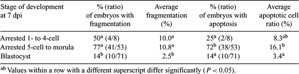
The relationship between embryonic fragmentation, embryonic arrest, and apoptosis has been the subject of some controversy (Hardy K 1999 Rev. Reprod. 4, 125–134). In order to investigate possible links, in vivo-produced, in vitro-cultured porcine embryos (n = 132) were scored for developmental stage and fragmentation at 7 days post insemination (dpi) and processed for propidium iodide and annexin V labelling. After fixation, embryos were processed for terminal deoxynucleotidyl transferase mediated dUTP nick end labelling (TUNEL). Using confocal microscopy, a cell was categorized apoptotic if (i) it had a fragmented or condensed nucleus, (ii) the cell membrane was annexin V-positive, and (iii) the nucleus was TUNEL labelled. An apoptotic cell ratio (ACR) was determined as the percentage of apoptotic cells per embryo. Differences in the % of fragmented and apoptotic embryos and correlations were analyzed using chi-square. Logistic regression was used to compare the average fragmentation % and the ACR. Sixty-one embryos (46%) arrested during the culture period, with 8 embryos arresting before or at the 4-cell stage. Significantly more arrested embryos were fragmented compared to embryos that were blastocysts at 7 dpi. Also, the average fragmentation percentage was significantly higher for arrested embryos compared to blastocysts. The correlation detected between developmental arrest and fragmentation was 0.60 (P < 0.05). None of the embryos without fragmentation had cells categorized as apoptotic, whereas 50 out of 55 embryos with fragmentation possessed apoptotic cells, which led to a correlation of 0.87 (P < 0.01) between fragmentation and apoptosis. The percentage of embryos with apoptotic cells was significantly higher for embryos arrested during the 5-cell to the morula stage compared to embryos that arrested before or at the 4-cell stage and embryos with blastocyst development at 7 dpi. The average ACR of embryos arrested during the 5-cell to the morula stage was significantly higher compared to the average ACR of blastocysts at 7 dpi. The correlation detected between the developmental arrest, during the 5-cell to the morula stage period and apoptosis was 0.57 (P < 0.01). Taken together, significant correlations between fragmentation, developmental arrest and apoptosis were detected. However, the association between embryonic arrest and apoptosis could be established only for embryos arrested after embryonic genome activation.
|