

318 ENHANCEMENT OF FERTILIZATION BY DIGITONIN IN ROUND SPERMATID INJECTION
S. Kishigami A , E. Mizutani A , S. Wakayama A and T. Wakayama AARIKEN, Center for Developmental Biology, Kobe, Japan. Email: kishigami@cdb.riken.jp
Reproduction, Fertility and Development 17(2) 309-310 https://doi.org/10.1071/RDv17n2Ab318
Submitted: 1 August 2004 Accepted: 1 October 2004 Published: 1 January 2005
Abstract
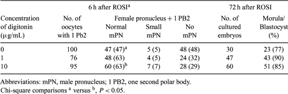
Reproductive technologies allow us to produce offspring using a variety of cells including sperm, spermatids, spermatocytes, somatic cells, and even parthenogenetic oocytes. In each of these technologies, failure of pronuclear formation after injection often prevents successful artificial reproduction. One of the possible causes is assumed to be that the breakage of the cytoplasmic membrane by simple pipetting is not enough to expose the nuclei to the ooplasm for pronuclear formation. To overcome this problem, we applied digitonin, a mild nonionic detergent, for the purpose of the permeabilization of cellular and nuclear membranes before injection. In this study, round spermatid cells in the mouse were used as a model because of their low pronuclear formation rate after injection. First, to examine the permeabilization of spermatids by digitonin, spermatid cells were incubated in CZB medium including 10 μg/mL of digitonin. Interestingly, the spermatids were lysed within 30 s after transfer but not other spermatogenic cells or somatic cells. Next, we conducted round spermatid injection (ROSI) using PVP including digitonin in a similar manner. Spermatids were picked up by injection pipette from spermatogenic cells suspended in a drop of PVP. These spermatids were transferred into another PVP drop including 1 μg/mL or 10 μg/mL of digitonin and left for 30 s. These digitonin-treated spermatids were then directly injected into previously activated oocytes. Six hours after injection, the fertilized oocytes were examined. Pronuclear formation rates were calculated as a proportion of oocytes with two pronuclei as well as one second polar body to total oocytes with one second polar body (Table 1). After digitonin treatment, fertilization rates significantly increased compared with ROSI without digitonin (Table 1). Further, these fertilized oocytes developed into blastocysts in vitro at comparable or higher rates. To further elucidate the effects of digitonin pretreatment on in vivo development, embryos were transferred into surrogate mothers 24 h after injection for offspring production. Although it is preliminary, we succeeded in the delivery of pups after ROSI with digitonin pretreatment (8 pups out of 14 transferred embryos). Thus, digitonin pretreatment is suggested to improve the success rate of ROSI.
|