

64 OOPLASMIC TRANSFER AFTER INTERSPECIES NUCLEAR TRANSFER: PRESENCE OF FOREIGN MITOCHONDRIA, PATTERN OF MIGRATION, AND EFFECT ON EMBRYO DEVELOPMENT
M. Sansinena A , J. Lynn B , R. Denniston A and R. Godke AA Embryo Biotechnology Laboratory, Reproductive Biology Center, Department of Animal Sciences, Louisiana State University
B Department of Biological Sciences, Louisiana State University, Baton Rouge, LA 70803, USA. Email: msansinena@agctr.lsu.edu
Reproduction, Fertility and Development 17(2) 182-182 https://doi.org/10.1071/RDv17n2Ab64
Submitted: 1 August 2004 Accepted: 1 October 2004 Published: 1 January 2005
Abstract
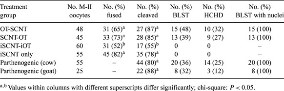
Interspecies somatic cell nuclear transfer (iSCNT) using the bovine cytoplast as universal recipient has potential applications in the conservation of exotic species. However, an in vitro developmental block has been observed using this approach. It has been suggested that mitochondrial mismatch between donor cell and recipient oocyte could be a cause for the embryonic developmental arrest. A series of experiments were conducted to investigate the effect of mixed mitochondrial populations (heteroplasmy) on early development of cloned embryos. In Experiment 1, we examined the effect of combining the technique of ooplasmic transfer (OT) with somatic cell nuclear transfer (SCNT) in the bovine model. In addition, presence and pattern of migration of foreign mitochondria after OT were examined by MitoTracker® (Molecular Probes, Inc., Eugene, OR, USA) staining. In Experiment 2, we examined the effect of transferring caprine ooplasm into bovine enucleated oocytes (iOT) used as recipients for goat iSCNT. Ooplasm from donor oocytes was aspirated until the oolema was ruptured and filled about 200 μm of the micropipette. Aspirated ooplasm was injected into recipient oocyte; the oolema of the recipient oocyte was also ruptured by partial aspiration into the micropipette to ensure mixing. Mean cleavage rates and embryo development were compared by chi-square analysis. Percentages (except for parthenogenic controls) were calculated from number of fused couplets. In Experiment 1, there was no significant effect of the sequence of events (OT-SCNT or SCNT-OT) on the number of fused, cleaved, blastocyst (BLST), or hatched blastocyst (HCHD) embryos (Table 1). MitoTracker Green FM staining of donor oocytes used for OT revealed foreign mitochondria were introduced by the procedure. Their pattern of distribution remained in a distinct cluster after 12, 74 and 144 h of in vitro culture. However, when goat ooplasm was injected into bovine enucleated oocytes used for iSCNT, there was a significant reduction in fusion (52 vs. 82%) and cleavage rates (55 vs. 78%) (P < 0.05). In addition, the procedure of iOT prior to iSCNT was not effective in overcoming the 8- to 16-cell in vitro developmental block and only parthenogenic cow and goat controls reached blastocyst (36 and 32%) and hatched blastocyst (25 and 12%) stages, respectively (Table 1). This study demonstrates that foreign mitochondria are introduced at the time of OT and these mitochondria remain in a cluster without relocation after a few mitotic divisions. Although the bovine cytoplast appears capable of supporting mitotic divisions after iOT-iSCNT, heteroplasmy or mitochondrial incompatibilities may affect nuclear-ooplasmic events occurring at genomic activation. To our knowledge, this is the first scientific report of iOT used in combination with iSCNT in an attempt to overcome the in vitro developmental block. Further research is needed to determine characteristics of foreign mitochondrial dynamics as well as replication of foreign mitochondria introduced into NT embryos.
|