413 ULTRASONOGRAPHIC EVALUATION OF FETAL LIVER SIZE DURING PREGNANCY IN BOVINE CLONED FETUSES
G. A. Jaúregui A , M. Panarace A , C. Garnil A , A. Segovia A , J. J. Lagioia A , J. Gutiérrez A , E. Rodriguez B , M. Marfil A and M. Medina AA Goyaike SAACIyF, Biotechnology Area, Carmen de Areco CC37, CP 6725, Buenos Aires, Argentina
B UNCPBA, Facultad de Ciencias Veterinarias, Tandil, Buenos Aires, Argentina
Reproduction, Fertility and Development 19(1) 322-322 https://doi.org/10.1071/RDv19n1Ab413
Submitted: 12 October 2006 Accepted: 12 October 2006 Published: 12 December 2006
Abstract
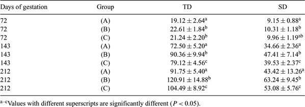
In ruminants, abnormal increased liver size is a common description cited in many postmortem studies performed on aborted fetuses and stillborn cloned calves (Heyman et al. 2002 Biol. Reprod. 66, 6–13). Although fetal liver size can be accurately determined throughout pregnancy using ultrasonography as a method of monitoring fetal health in humans, there are no reports of this being done in cattle. Thus, the aim of this study was to prospectively characterize the ultrasonographic fetal liver growth pattern in IVF-derived pregnancies, and then to establish comparisons with measurements taken from cloned fetuses. For this purpose, (A) IVF-derived pregnancies were used as the control group (n = 10), and the cloned ones were split into 2 groups according to the outcome of their pregnancies: (B) clones that died between 110 and 282 days (n = 21), and (C) clones born alive (n = 16). All recipients were multiparous, nonlactating, and Aberdeen Angus breed. Measurements were done by ultrasonography (Toshiba Nemio 20, Tokyo, Japan) using a 5–10 MHz intraoperative finger probe from 72 to 114 days (transrectal) and a 3–6 MHz linear-array probe between 143 and 212 days (transabdominal). Three parameters of liver size were measured: (a) in a coronal (longitudinal) plane of the fetus, cephalocaudal diameter (CC) was taken from the dome of the right hemi-diaphragm to the tip of the right lobe; (b) in an axial (transverse) plane at right angles to one another passing through or slightly caudal to the portal umbilical venous complex: (b1) transverse diameter (TD) and (b2) sagittal diameter (SD) were determined (Gimondo et al. 1995 J. Ultrasound Med. 14, 327–333). A repeated-measure ANOVA detected significant interaction for the 3 variables included in this study (P < 0.01); therefore, differences between groups at each week of gestation were analyzed using the Wald-Wolfowitz test (InfoStat V1.5; FCA, Universidad de Córdoba, Córdoba, Argentina). Results showed that CC, TD, and SD were statistically smaller in the IVF group throughout pregnancy (P < 0.05). No significant differences between (B) and (C) groups were found at 72 and 87 days (P > 0.05). However, from 143 days onward, liver of fetuses from the (B) group were statistically larger in CC, TD, and SD diameters than those values obtained in the (A) and (C) groups (P < 0.05) (Table 1). To conclude, the present study showed that fetal liver size can be measured throughout gestation in cows using noninvasive ultrasonography. Secondly, since fetuses from (A) and (C) groups were born alive, increased abnormal size measurements after 140 days can be used as indicator of clones that fail late in gestation (B). Also, this methodology may enhance sonographic assessment of fetal growth abnormalities and conditions with fetal liver involvement. Further studies will be necessary to evaluate differences between naturally conceived and IVF fetuses and the correspondence of this three-dimensional evaluation of the liver and to estimate its weight.

|


