256 EFFECT OF EPIDERMAL GROWTH FACTOR ON IN VITRO MATURATION OF CYNOMOLGUS MONKEY (MACACA FASCICULARIS) OOCYTES
J. Yamasaki A , J. Okahara-Narita A , C. Iwatani A , H. Tsuchiya A , S. Nakamura A , N. Sakuragawa B and R. Torii AA Research Center for Animal Life Science, Shiga University of Medical Science, Otsu, Shiga, Japan;
B Department of Regenerative Medicine, School of Allied Health Sciences, Kitasato University, Sagamihara, Kanagawa, Japan
Reproduction, Fertility and Development 20(1) 208-208 https://doi.org/10.1071/RDv20n1Ab256
Published: 12 December 2007
Abstract
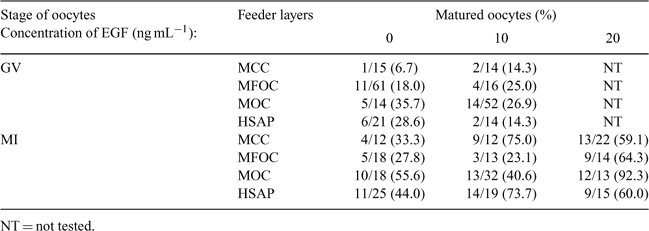
Collected oocytes include not only mature oocytes (metaphase II: MII), but also immature oocytes (germinal vesicle: GV, and metaphase I: MI). To establish a dependable artificial indoor breeding program in cynomolgus monkeys, we are planning to carry out in vitro maturation (IVM) using GV and MI oocytes. In this study, we attempted to determine whether different types of feeder layers and epidermal growth factor (EGF) were effective for IVM. Cumulus–oocyte complexes (COCs) were collected from ovaries of 4–10-year-old female cynomolgus monkeys stimulated by the combination of FSH (25 IU kg–1 × 9 days) and hCG (400 IU kg–1) (Torii 2000 Primates 39, 399–406). Oocytes were classified by morphological features: oocytes retaining an intact germinal vesicle nucleus (GV); oocytes that had undergone germinal vesicle breakdown without polar body formation (MI); and oocytes with a first polar body (MII). GV and MI oocytes were co-cultured on monkey cumulus cells (MCC), monkey follicular ovarian cells (MFOC), monkey oviductal cells (MOC), or human solubilized amnion product (HSAP), with TCM-199+10% fetal bovine serum containing epidermal growth factor (EGF; 10 ng mL–1 or 20 ng mL–1). The maturation rate from GV to MII oocytes was 6.7% (MCC), 18.0% (MFOC), 35.7% (MOC), and 28.6% (HSAP) (Table 1). Although higher maturity was observed in MOC and HSAP, the effect of EGF was not found in co-cultures using any feeder layers. The maturation rate from MI to MII oocytes was 33.3% (MCC), 27.8% (MFOC), 55.6% (MOC), and 44.0% (HSAP) (Table 1). The highest maturation rate from GV and MI was observed in co-cultures using MOC. The maturation rate from MI to MII oocytes in the presence of 10 ng mL–1 EGF was 75.0% (MCC) and 73.7% (HSAP) (Table 1), whereas the rate in the presence of 20 ng mL–1 EGF was 59.1% (MCC), 64.3% (MFOC), 92.3% (MOC), and 60.0% (HSAP) (Table 1). Thus, the best maturation rate was a co-culture using MOC as a feeder layer with 20 ng mL–1 EGF. According to our results, maturation rate during IVM depends on the cellular type of feeder layers and the concentration of EGF. EGF is especially effective for maturity from MI to MII oocytes, but not from GV to MI or MII oocytes. Thus, IVM should be carred out under optimal culture conditions, including suitable feeder layer and media plus supplements. In the future, it is important that intracytoplasmic sperm injection be carried out using in vitro-matured MII oocytes for establishment of an artificial indoor breeding program in cynomolgus monkeys.

|


