

309 PRODUCTION OF CATTLE BY NUCLEAR TRANSFER FROM CELLS IN WHICH A GENE IS DISRUPTED
M. Urakawa A , Y. Sendai B , A. Ideta A , K. Hayama A , Y. Shinkai C , T. Sawada D , H. Hoshi E and Y. Aoyagi AA ET Center, ZEN-NOH, Kamishihoro, Hokkaido, Japan;
B Central Research Institute for Feed and Livestock, ZEN-NOH, Tsukuba, Ibaraki, Japan;
C Institute for Virus Research, Kyoto University, Kyoto, Japan;
D Second Department of Surgery, Dokkyo University School for Medicine, Mibu, Tochigi, Japan;
E Research Institute for Functional Peptides, Yamagata, Japan
Reproduction, Fertility and Development 21(1) 251-252 https://doi.org/10.1071/RDv21n1Ab309
Published: 9 December 2008
Abstract
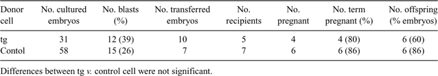
Gene-targeted animals provide a powerful model to examine gene functionality. In this study, we examined the effect of gene targeting of donor cells for nuclear transfer (NT) on the pregnancy rate and on viability of the offspring after embryo transfer. Gene-targeted (tg; targeting of both alleles of the gene encoding bovine prion protein) or non-manipulated (control) bovine fetal fibroblasts were used for NT. A promoterless positive selection vector (pPrP5.2) containing an internal ribosome entry site-antibiotic resistance gene (neo) cassette and loxP sequences was used to disrupt the bovine prion protein gene. The cells (tg) in which homologous recombination was occurred were used for NT. The tg and control cells were cultured in DMEM with 10% FCS and were prepared in the early G1 phase to our previous report (Urakawa M et al. 2004 Theriogenology 62, 714–728). Each donor cell was inserted into an enucleated in vitro-matured (19 h) oocyte. Cell fusion (DC, 200 V mm–1, 10 μs) and activation (DC, 100 V mm–1 , 60 μs) were done in 0.3 m mannitol solution. The NT embryos were then activated with 5 μm Ca-ionophore and 10 μg of mL–1 cycloheximide and were cultured with bovine oviduct epithelial cells in CR1aa with 5% CS. The blastocyst rates were judged at 6 days after NT. The blastocysts were non-surgically transferred to recipient heifers. The recipients were monitored daily for heat behavior, examined by ultrasound at Day 30 and 60, and then observed monthly to confirm pregnancy. The offspring born in the tg group were confirmed by PCR to be transgenic. Statically significance was tested using a chi-square test or t-test. Developmental rate to the blastocyst stage, pregnancy rate at Day 30 and 60, and calving rate did not differ significantly between tg and the control group (Table 1). Gestation length (tg; 290.0 ± 2.2 days v. control; 290.5 ± 3.9 days) and birth weight (tg; 39.6 ± 8.0 kg v. control; 40.2 ± 4.1 kg) were not significantly different. These results indicate that gene targeting of donor cells used for NT does not significantly affect the development of embryos, pregnancy rate, or the viability of the offspring.
|