

17 TREATMENT WITH MGCD 0103 IMPROVES THE IN VITRO DEVELOPMENT OF PORCINE EMBRYOS DERIVED FROM SOMATIC CELL NUCLEAR TRANSFER
H.-Y. Zhu A , L. Jin A , Q. Guo A , Y.-C. Zhang A , X.-C. Li A , J.-D. Kang A and X.-J. Yin AYanbian University Jilin Provincial Key Laboratory of Transgenic Animal and Embryo Engineering, Yanji, Jilin, China
Reproduction, Fertility and Development 28(2) 138-139 https://doi.org/10.1071/RDv28n2Ab17
Published: 3 December 2015
Abstract
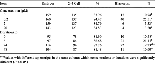
We use MGCD 0103 to test whether the treatment with this novel histone deacetylase inhibitor improves the in vitro development of porcine somatic cell NT (SCNT) embryos. Matured eggs were cultured in medium supplemented with 0.05 M sucrose and 0.4 μg mL–1 demecolcine for 1 h. Treated eggs with a protruding membrane were transferred to medium supplemented with 5 μg mL–1 cytochalasin B and 0.4 μg mL–1 demecolcine. Protrusions were then removed by aspirating with a 15-μm inner diameter glass pipette. A single donor cell was inserted into the perivitelline space of each egg and electrically fused using 2 direct pulses of 150 V mm–1 for 50 μs in 0.28 M mannitol. Fused eggs cultured for 1 h were activated by 2 direct pulses of 100 V mm–1 for 20 μs and incubated with 2 mM 6-DMAP for 4 h. Subsequently, the cloned embryos were cultured in medium for 7 days at 38.5°C in 5% CO2 humidified air. In Experiment 1, after activation and treatment with 6-DMAP for 4 h, the SCNT embryos were cultured in medium supplemented with 0, 0.2, 2, or 20 μM MGCD 0103 for 24 h and then transferred to medium without MGCD 0103. In Experiment 2, SCNT embryos were cultured in medium supplemented with 0.2 μM MGCD 0103 for 0, 6, 24, or 48 h and then transferred to medium without MGCD 0103. As shown in Table 1, development to the blastocyst stage increased in SCNT embryos treated with 0.2 μM MGCD 0103 compared with the control or groups treated with 2 or 20 μM MGCD 0103 (25.51 v. 10.74, 3.53, 3.20%, respectively; P < 0.05). As shown in Table 1, treatment for 6 h with 0.2 μM MGCD 0103 significantly improved the rate of blastocyst formation compared with the control or groups treated for 24 or 48 h (21.17 v. 10.48, 19.23, 10.20%, respectively; P < 0.05). Our results suggested that 0.2 μM MGCD 0103 treatment for 6 h can improve in vitro developmental competence of porcine SCNT embryos.
|