

73 APPLICATION OF THE HOLLOW FIBER VITRIFICATION METHOD TO THE CRYOPRESERVATION OF HIGHLY CRYOSENSITIVE EMBRYOS
A. Uchikura A , H. Matsunari A B , K. Nakano A , S. Hatae A , Y. Matsumura A , Y. Asano A , T. Takeishi A , H. Nakauchi C and H. Nagashima A BA Laboratory of Developmental Engineering, Department of Life Sciences, School of Agriculture, Meiji University, Kawasaki, Kanagawa, Japan;
B Meiji University International Institute for Bio-Resource Research (MUIIBR), Kawasaki, Kanagawa, Japan;
C Institute of Medical Science, University of Tokyo, Minato, Tokyo, Japan
Reproduction, Fertility and Development 27(1) 129-130 https://doi.org/10.1071/RDv27n1Ab73
Published: 4 December 2014
Abstract
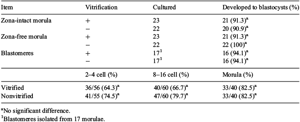
We recently demonstrated that the hollow fibre vitrification (HFV) method (Matsunari et al. 2012) could effectively be applied to the cryopreservation of embryos from diverse species. In this study, we applied the HFV method to the cryopreservation of highly cryosensitive specimens, such as in vitro matured (IVM)/IVF-derived porcine zona-free morulae and blastomeres isolated from those morulae, as well as IVM/IVF-derived cattle embryos at early cleavage stages. Porcine parthenogenetic morulae (d-4) derived from IVM oocytes were treated with 0.25% pronase to remove zona pellucidae. The resulting blastomeres were isolated from the zona-free morulae by a decompaction treatment followed by gentle pipetting. Bovine IVM-IVF embryos at the 2 to 4 cell (d-1), 8 to 16 cell (d-3), and morula stages (d-5) were then subjected to vitrification. The HFV procedure was performed as described previously using 15% dimethyl sulfoxide, 15% ethylene glycol, and 0.5 M trehalose as cryoprotectants. Four to twenty embryos, or all of the blastomeres isolated from a single morula, were individually loaded into a cellulose acetate hollow fibre (25 mm long, 185 μm φ, 15 μm membrane thickness) and vitrified. Survival of the vitrified embryos was assessed by in vitro development to blastocysts. Blastomeres recovered after vitrification were aggregated in micro-wells to examine their ability to form blastocysts. The HFV method was demonstrated to be effective for cryopreserving zona-free in vitro-produced porcine morulae and the blastomeres isolated from them (Table 1), as well as bovine IVM-IVF embryos at early cleavage stages. These data demonstrate that the HFV method is effective for highly cryosensitive specimens, such as IVM/IVF-derived porcine zona-free morulae and blastomeres isolated from those morulae, and IVM/IVF-derived cattle embryos at early cleavage stages. These achievements may expand the technological options in the production of cloned and genetically modified pigs that are useful for biomedical research.
|
This study was supported by JST, ERATO, the Nakauchi Stem Cell and Organ Regeneration Project, and MUIIBR.