34 LIFESPAN AND CHROMOSOMAL STABILITY OF BOVINE AND PORCINE FETAL FIBROBLAST CELLS CULTURED IN VITRO
A.M. Giraldo A C , J.W. Lynn B , C.E. Pope C , R.A. Godke A and K.R. Bondioli AA Department of Animal Sciences, Louisiana State University, Baton Rouge, LA 70803, USA
B Department of Biological Sciences, Louisiana State University, Baton Rouge, LA 70803, USA
C Audubon Institute for Research of Endangered Species, New Orleans, LA 70148, USA. Email: agiral1@lsu.edu
Reproduction, Fertility and Development 17(2) 167-167 https://doi.org/10.1071/RDv17n2Ab34
Submitted: 1 August 2004 Accepted: 1 October 2004 Published: 1 January 2005
Abstract
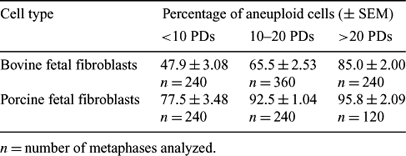
The low efficiency of nuclear transfer (NT) has been related to factors such as mitochondria heteroplasmy, failure of genomic activation, and asynchrony between the donor karyoplast and recipient cytoplast. Few studies have characterized donor cell lines in terms of proliferative capacity and chromosomal stability. It is known that suboptimal culture conditions can induce chromosomal abnormalities, and the use of aneuploid donor cells during NT can lead to a high incidence of abnormal cloned embryos (Giraldo et al. 2004 Reprod. Fertil. Dev. 16, 124 abst). The purpose of this study was to determine the lifespan and chromosomal stability of bovine and porcine fetal cells. Four bovine and four porcine fibroblast cells lines were established from 50-day and 40-day fetuses, respectively. Cells were cultured in DMEM medium supplemented with 10% fetal bovine serum and 1% penicillin and streptomycin at 37°C in 5% CO2. Each cell line was passaged to senescence. Total population doublings (PDs) and cell cycle duration were calculated. To determine the chromosome numbers at different PDs, cells were synchronized in metaphase, fixed, and stained. ANOVA and chi-square tests were used to analyze differences in PDs and proportion of aneuploid cells between cell lines, respectively (P < 0.05). The results show that proliferative capacity was not different between cell lines derived from the same species. Cell lines derived from bovine and porcine fetuses had different in vitro lifespans (33 PDs vs. 42 PDs, respectively; P < 0.05). The mean length of the cell cycles for both bovine and porcine fetal fibroblasts was ∼28 h. The percentage of aneupliod cells in both bovine and porcine fetal cell lines increased progressively with duration of culture (see Table) and was high throughout the study. The proliferative capacity of cultured cells was similar within individuals of the same species, but growth characteristics differed between fetal bovine and porcine cell lines. The progressive increase of aneuploid cells could be due to suboptimal culture conditions or unusual chromosome instability in the particular fetuses used. These data demonstrate the importance of determining chromosome content and the use of cells at early passages to decrease the percentage of aneuploid reconstructed embryos and increase the efficiency of NT.

|


