

183 DEVELOPMENT OF PORCINE EMBRYOS MICROINJECTED WITH PORCINE EMBRYONIC GERM CELLS
C.-H. Park A B , S.-G. Lee A B , D.-H. Choi A B , M.-G. Kim A B and C. K. Lee A BA School of Agricultural Biotechnology, Seoul National University, Seoul, South Korea
B Xenotransplantation Research Center, Seoul National University Hospital, Seoul, South Korea
Reproduction, Fertility and Development 18(2) 199-199 https://doi.org/10.1071/RDv18n2Ab183
Published: 14 December 2005
Abstract
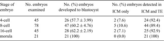
Embryonic germ (EG) cells, derived from primordial germ cells in the developing fetus, are similar to embryonic stem (ES) cells in terms of expression pattern of undifferentiated markers and their ability to colonize both the somatic and the germ cell lines following injection into a host blastocyst, which has been proven in mouse. Several studies using porcine EG cells have shown that it is possible to produce somatic chimeras after blastocyst injection. However, not only was the degree of reported chimerism low, but also there has been no report about the fate of injected EG cells in porcine blastocysts. This study was designed to observe the distribution pattern of porcine EG cells in chimeric blastocyst after injection into cleavage-stage porcine embryos. To ascertain development of microinjected porcine embryos with EG cells, 10 to 15 EG cells were injected into cleavage stage of in vitro fertilized embryos and cultured up to blastocyst. Also, porcine EG cells were labeled with DiO (Invitrogen, Carlsbad, CA) on the cell membrane or transfected with green fluorescent protein gene to observe whether the EG cells injected in the host embryo would incorporate into the inner cell mass (ICM) or trophectoderm (TE). Chimeric embryos were produced and allowed to develop into blastocysts to investigate the injected EG cells would come to lie in ICM and/or TE of the blastocyst, by scoring their position. In result, developmental rate was similar in all treatments. In all treatments, EG cells were mainly allocated in both ICM and TE of the chimeric blastocysts. These results suggest that examining the allocation pattern of injected EG cells, maintained pluripotency in vitro, could provide clues of differentiation process in vivo. Furthermore, to enhance the allocation of EG cells into the embryonic lineage, it would be required to optimize the culture condition for EG cells as well as embryos. Further experiment are needed to determine whether the injected EG cells could maintain their properties throughout the environment in the embryonic development in vitro.
|