

123 PROGESTERONE LEVELS DURING 20 DAYS OF PREGNANCY IN RABBIT TREATED FOR ENDOMETRIOSIS OR WITH ANTI-CD44
M.J. Illera A , P. Bermejo A , A. Natarajan A , C. Willingham A and J. Hernandez AAFacultad de Veterinaria, Universidad Complutense de Madrid, Madrid, Spain. Email: mjillera@vet.ucm.es
Reproduction, Fertility and Development 17(2) 212-212 https://doi.org/10.1071/RDv17n2Ab123
Submitted: 1 August 2004 Accepted: 1 October 2004 Published: 1 January 2005
Abstract
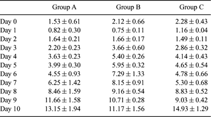
Embryo implantation begins when the blastocyst both assumes a fixed position in the uterus and establishes a more intimate relationship with the endometrium. Successful implantation depends upon hormonal synchronization and development and the receptivity of the endometrium. CD44 is a cell surface molecule that has been implicated in the initial attachment of the embryo. The aim of this work was to study the hormonal levels of P4 in three groups of animals which have a normal pregnancy or an induced reduction in the number of implants. Twelve adult New Zealand does (n = 12) were naturally inseminated with a buck of proven fertility. Blood samples were obtained daily during 20 days of pregnancy. Hormonal determinations were performed by enzyme immunoassay. Animals were divided into three groups: group A (n = 4): control animals; group B (n = 4): endometriosis was surgically induced in the right horn a month before the animal was mated; and group C (n = 4): animals received an injection of 20 micrograms of anti-CD44 in the right horn via mid-ventral laparotomy on Day 6.5 post-coitum (0.5 mL each, from the ovarian end to the cervix). Each animal served as her own control with the left uterine horn receiving 0.5 mL of saline. Statistical analysis was performed using ANOVA.The number of corpora lutea was similar in all treatments. No statistical differences were found comparing CLs in the right/left ovary. In group A, a mean of 3.4 ± 0.47 (mean ± SE) implants was found in the right horn while the mean in the left side was 4.6 ± 0.81. In group B, a marked reduction in implantation sites was found, with 1.8 ± 0.60 and 4.66 ± 0.84 on the right and left horns, respectively. With anti-CD44 injected into the uterine horn (Group C), a mean of 0.12 implant was present in the right uterine horn compared with 3.6 implants on the left side (P < 0.001). Progesterone levels from Days 1 to 10 are shown in the following table (mean ± SE). Comparisons in day values are not statistically significant P > 0.05. After Day 10 the levels of progesterone were similar in all groups. The results showed an increase of progesterone levels in group B; this could be due to endometriosis and not to the number of implants. The results in the CD44 group reveal that progesterone profiles were similar to those in the control group, and we can conclude that the reduced number of implants found in group C did not affect the progesterone levels.
|