

201 EFFECT OF HEPARIN AND CALCIUM IONOPHORE ON ACROSOME REACTION IN EPIDIDYMAL SPERMATOZOA OF DROMEDARY CAMEL (CAMELUS DROMEDARIUS)
N.A. Wani A and M.A. Nowshari AACentral Veterinary Research Laboratory, Dubai, United Arab Emirates. Email: mnowshari@hotmail.com
Reproduction, Fertility and Development 17(2) 250-251 https://doi.org/10.1071/RDv17n2Ab201
Submitted: 1 August 2004 Accepted: 1 October 2004 Published: 1 January 2005
Abstract
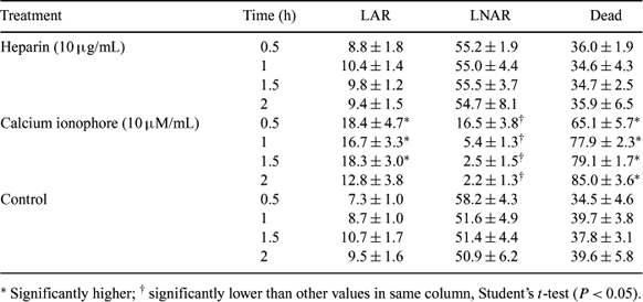
The present study was undertaken to evaluate the effect of heparin and calcium ionophore (Ca-I) on the induction of acrosome reaction in epididymal spermatozoa of dromedary camel. Cauda epididymides were isolated from testes of mature males immediately after slaughter at a local abattoir and brought to laboratory in saline solution on ice (0–1°C). Spermatozoa were collected by aspirating the sperm-rich fluid from convoluted tubules beneath the ductus deferens with a sterile hypodermic needle attached to a 10-mL syringe containing 2–4 mL of TRIS-tes egg yolk diluent and stored overnight at 4°C. The sperm suspension was aliquoted into 3 tubes, washed twice with fertilization medium (TALP; Parrish et al. 1985 Theriogenology 24, 537); pellets were dissolved in 1 mL of fertilization medium supplemented with 10 μg/mL heparin or 10 μM/mL Ca-I or no additive (control). Aliquots (200 μL) were made in Eppendorf tubes and incubated at 38°C for 0.5, 1, 1.5, or 2 h. Sperm viability and acrosomal status was evaluated by a dual staining procedure (Didion et al. 1989 Gamete Research 22, 51–57). Briefly, spermatozoa were incubated with an equal volume of 0.2% trypan blue for 10 min and washed twice (centrifugation at 700g for 6 min) with fertilization medium. Smears were made on glass slides and dried quickly on a warm stage. Slides were stained with 10% giemsa stain for 40 min. They were rinsed under a stream of distilled water, air-dried, and covered with DPX mountant and coverslips. The experiment was replicated six times. Spermatozoa (n > 200/slide) were examined in randomly selected microscopic fields under a phase-contrast microscope at 1000×. Spermatozoa were classified as dead (stained blue in the post-acrosomal region), live non-acrosome-reacted (unstained post acrosomal region but light purple-dark pink acrosome) or live acrosome reacted (unstained acrosome and post-acrosomal region). Data were analyzed using Student's t-test and are presented as mean percent ± SEM. There was no difference in the proportion of live acrosome-reacted (LAR) and live non-acrosome-reacted (LNAR) spermatozoa in the heparin group compared to the control group (Table 1). Although Ca-I group had higher LAR sperm, the proportion of dead spermatozoa was high (P < 0.05). It may be concluded that heparin was not able to enhance the acrosome reaction, and Ca-I seems to be detrimental to the spermatozoa at this concentration. Further investigations are warranted to evaluate the appropriate concentrations and duration of exposure to both of these agents for induction of acrosomal reaction in spermatozoa of this species.
|