

203 PRODUCTION OF ANDROGENETIC SHEEP BLASTOCYSTS
K. Matsukawa A B , M. Y. Turco A , G. Ptak A and P. Loi AA Department of Comparative Biomedical Sciences, University of Teramo, Teramo, Italy
B National Institute of Livestock and Grassland Science, Tsukuba, Ibaraki, Japan
Reproduction, Fertility and Development 19(1) 218-219 https://doi.org/10.1071/RDv19n1Ab203
Submitted: 12 October 2006 Accepted: 12 October 2006 Published: 12 December 2006
Abstract
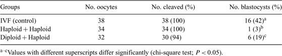
The androgenetic embryo is a useful model for investigating the contribution of the paternal genome (e.g. genomic imprinting) to embryonic development. Few works on androgenetic embryo production in domestic animals exist. In this study, we compared the developmental ability of diploid, haploid, and triploid androgenetic sheep embryos. In vitro-matured metaphase II oocytes were enucleated in HEPES-buffered TCM-199 with cytochalasin B (7.5 µg mL−1) and Hoechst 33342 (5 µg mL−1) under UV light using a Narishighe Micromanipulator fitted to an inverted Nikon microscope. Enucleated oocytes were fertilized in vitro with a high sperm concentration (2.5 × 10⁷ sperm mL−1). Fifteen hours after in vitro fertilization (IVF), embryos were centrifuged (12 000g for 10 min) to visualize the pronuclei; the number of pronuclei were scored under the inverted microscope. In Experiment 1, IVF (control), haploid (1 pronucleus), diploid (2 pronuclei), and triploid (3 pronuclei) embryos were cultured in SOFaa medium with BSA, according to the protocol in our laboratory (Ptak et al. 2002 Biol. Reprod. 67, 1719–1725). In Experiment 2, we performed pronuclear transfer to produce diploid embryos. A single pronucleus was aspirated with a bevelled pipette from haploid (haploid + haploid) or diploid (diploid + haploid) embryos and transferred into the perivitelline space of another haploid embryo. The reconstructed zygotes were electrofused in 0.27 M mannitol solution with 50 µM CaCl2 and 100 µM MgCl2 by a single DC pulse (0.8 kV cm−1 for 80 µs). As a control group for Experiment 2, IVF embryos (pronuclear stage) were centrifuged, followed by the aspiration of a small volume of cytoplasm, and fused under the same condition of diploidization. In Experiment 1, there was no significant difference in cleavage rate (91% to 98%), but there was a significant difference on blastocyst development between IVF and androgenetic embryos (IVF: 43% (26/60); haploid: 0% (0/37); diploid: 1% (1/73); and triploid: 2% (1/48)). In Experiment 2, there was no significant difference in cleavage rate (94% to 100%). However, there was a significant difference on blastocyst development (Control: 42%; haploid + diploid: 19%; and haploid + haploid: 3%; Table 1). Our results suggest that sheep androgenetic embryos show poor developmental ability compared with IVF embryos. Interestingly, diploid androgenetic embryos produced by IVF displayed very poor development; however, such poor development was rescued, for unknown reasons, by pronuclear transfer. Ongoing experiments will provide new insight into this previously uncharacterized phenomenon. In conclusion, pronuclear transfer was an effective method for producing sheep androgenetic blastocysts in vitro.
|