

45 DEVELOPMENT OF BOVINE NUCLEAR TRANSFER EMBRYOS DERIVED FROM ADULT MARROW STROMAL CELLS AND FETAL MUSCLE CELLS
M. Murakami A , X. J. Bai A B , W. S. Shi B , W. M. Wang B , W. Liu B and Y. J. Dong A BA Animal Embryo Engineering Center, Qingdao Agriculture University, Qingdao, Shandong, China;
B Shandong Provincial Research Institute for Black Cattle Breed Engineering, Qingdao, Shandong, China
Reproduction, Fertility and Development 21(1) 122-123 https://doi.org/10.1071/RDv21n1Ab45
Published: 9 December 2008
Abstract
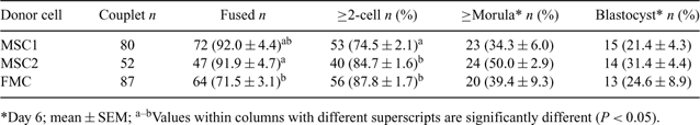
The use of less differentiated cells, such as marrow stromal cells (MSCs), as the nuclear donor may increase the efficiencies of somatic cell cloning in cattle. Healthy offspring was produced from bovine MSCs (Kato et al. 2004 Biol. Reprod. 70, 415–418); however, there is little information that directly compared the post-implantation survival among the clones originated from MSCs and other somatic cells. The objective of this study was to evaluate the developmental potential in vitro and in vivo of bovine NT embryos derived from adult MSCs and fetal muscle cells (FMCs). Primary cell populations of MSCs and FMCs were obtained from the femurs of 8- and 12-months-old Holstein cows (MSC1 and MSC2 groups, respectively) and a Holstein fetus at 8 months of gestation (FMC group), respectively. They were used as donor cells for the NT procedure (Murakami et al. 2005 Cloning Stem Cells 7, 77–81) at passages 1 to 3. Briefly, oocytes collected from cow ovaries were enucleated at 20 h post-in vitro maturation (IVM), and the donor cell was placed into the perivitelline space. The couplets were fused electrically, activated (10 μg mL–1 cycloheximide; 4 h), and cultured in CR1aa medium. Development in vitro of these embryos is summarized in Table 1. Data were analyzed by ANOVA. The fusion rates were higher in the MSC groups than in the FMC group. The rate of cleaved embryos was significantly lower (P < 0.05) in the MSC1 group than in the other groups. However, there were no significant differences among the groups in the rates of development into morulae/blastocysts on Day 6. A total of 8 and 3 fresh good quality Day 6 embryos in the MSC1 and FMC groups, respectively, were nonsurgically transferred to 6 naturally cycling Holstein females 6 days after estrus (3 recipients/group, 1–3 embryos/female). On Day 30 of gestation, none of the recipients were pregnant in the FMC group, while 2 recipients in the MSC1 group were diagnosed as pregnant via ultrasonography; they remained pregnant on Day 80 of gestation. In addition, a total of 4 Day 7 embryos cryopreserved in 1.8 m ethylene glycol plus 0.05 m trehalose were directly transferred to 4 synchronized recipients after thawing (1 embryo/female) in the MSC1 group. Of those, 2 females were pregnant on day 30 of gestation. These results indicate that the developmental potential in vitro of bovine NT embryos derived from adult MSCs was comparable to that of the embryos derived from fetal muscle cells, and that pregnancies were produced after transfer of the fresh and frozen–thawed NT embryos derived from the MSC, but the sample size was small. Further studies with more replicates are needed to evaluate viability in vivo of these cloned embryos for comparative purposes.
|