First record of Pseudocercospora mori causing grey leaf spot on mulberry in Australia
K. R. E. Grice A D , V. Beilharz B and R. G. Shivas CA Centre for Tropical Agriculture, Department of Primary Industries and Fisheries, 28 Peters Street, Mareeba, Qld 4880, Australia.
B Institute for Horticultural Development, Department of Primary Industries, Private Bag 15, Ferntree Gully Delivery Centre, Vic. 3156, Australia.
C Plant Pathology Herbarium, Department of Primary Industries and Fisheries, 80 Meiers Road, Indooroopilly, Qld 4068, Australia.
D Corresponding author. Email: kathy.grice@dpi.qld.gov.au
Australasian Plant Disease Notes 1(1) 9-10 https://doi.org/10.1071/DN06005
Accepted: 17 July 2006 Published: 24 August 2006
Abstract
Grey leaf spot of mulberry caused by Pseudocercospora mori is reported for the first time from Australia. It was identified on both white mulberry (Morus alba) and black mulberry (Morus nigra).
Pseudocercospora mori (Hara) Deighton, the grey leaf spot pathogen of mulberry, has been previously reported from Japan, Democratic Republic of the Congo, Taiwan, China, the USA (Chupp 1954) and India (Srivastava et al. 1978). The fungus can cause significant damage to leaves of Morus spp. causing necrotic spots, which become brittle, resulting in shot hole, leaf yellowing and defoliation (Babu et al. 2002).
In June 2003, samples of prematurely senescing leaves of white mulberry (Morus alba L.) and black mulberry (Morus nigra L.) were collected from two separate properties (non-commercial), both in the Daintree area of north Queensland. Symptoms on both samples appeared as a blotch with conidia being abundantly produced on the underneath surface of the leaves.
The fungus on the infected mulberry leaves was identified as Pseudocercospora mori by comparison with the descriptions and illustrations in Hsieh and Goh (1990) and Babu et al. (2002).
Pseudocercospora mori (Hara) Deighton. Mycological Papers 140: 148 (1976)
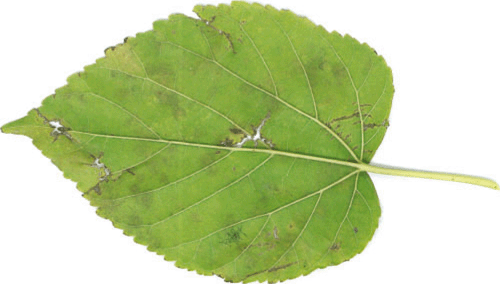
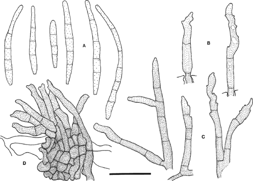
Leaf spots indistinct on upper surface, more conspicuous on the lower surface where they are brown, angular or spreading (particularly along leaf margins) and restricted by the larger veins, up to 7 mm wide (Fig. 1). Sporulation hypogenous, ranging from olivaceous and velutinous to darker brown and punctate. Immersed hyphae pale olivaceous, smooth, septate, branching, 1–3 µm diam. Superficial hyphae sparse, emerging from stomata, pale olivaceous, smooth, septate, 1–2 µm diam., bearing lateral conidiophores similar to those associated with stomatal egress. Stromata lacking, reduced to a few light brown cells in the substomatal cavity, or compact and light brown, filling the substomatal cavity, 18–25 µm diam. Conidiophores solitary or 2–22 in a divergent fascicle, olivaceous brown throughout or slightly paler at the apex, irregular in width, branched, straight, curved or sinuous, markedly geniculate towards a conic apex, conspicuously 1–4(–7) septate, 30–67 µm long × 3–4.5 µm wide. Conidiogenous loci not darkened, not thickened, non-refractive, 0.5–1 µm diam. Conidia pale olivaceous, straight or curved, smooth, narrowly obclavate, straight to mildly curved, occasionally constricted, usually narrowing gradually to a subobtuse or obtuse apex, and a little more abruptly to an obconic, truncate base, 1–5(–7) septate, 18–61× 3–4 µm (Fig. 2). Hila not darkened, not thickened, non-refractive, 1–2 µm diam.

|

|
Hsieh and Goh (1990) recorded conidiophores measuring 20–90 × 3–5 µm and conidia measuring 20–80 × 3–5 µm. They did not report the presence of external hyphae, but this feature is known to be variable and strongly influenced by the prevailing microclimate in several species of Pseudocercospora.
Specimens have been deposited in the Department of Primary Industries and Fisheries Plant Pathology Herbarium as BRIP 39897 and BRIP 39946. Since the initial findings, two other specimens of M. nigra collected near Walkamin on the Atherton Tableland (north Queensland) in July 2003 were also confirmed as Pseudocercospora mori.
This is the first record of Pseudocercospora mori in Australia. It is not considered to be of quarantine significance as diseases of mulberry are primarily of economic importance in silk producing countries where mulberry is the sole food plant of the silkworm.
Babu AM,
Kumar V, Govindaiah
(2002) Surface ultrastructural studies on the infection process of Pseudocercospora mori causing grey leaf spot disease in mulberry. Mycological Research 106, 938–945.
| Crossref | GoogleScholarGoogle Scholar |

Srivastava RC,
Khan MS, Lal MP
(1978) Some new leaf spot fungi from India. Indian Phytopathology 31, 524–525.


