

Leaf spot of cultivated and wild Alstroemeria spp. caused by Asperisporium alstroemeriae
S. Wolcan A B C , C. Rollán A and L. Ronco AA Centro de Investigaciones de Fitopatología, Facultad de Ciencias Agrarias y Forestales, UNLP, 60 y 119, La Plata 1900, Buenos Aires, Argentina.
B Comisión de Investigaciones Científicas de la Provincia de Buenos Aires, 526 e/10 y 11, La Plata 1900, Buenos Aires, Argentina.
C Corresponding author. Email: swolcan@speedy.com.ar
Australasian Plant Disease Notes 1(1) 33-35 https://doi.org/10.1071/DN06015
Accepted: 15 October 2006 Published: 24 October 2006
Abstract
Asperisporium alstroemeriae (Allesch.) Maubl., the causal agent of leaf spot on Alstroemeria spp., is recorded for the first time in Argentina on commercial hybrids and on the native species A. psittacina. It appears that cultivated hybrids of Alstroemeria spp. and A. psittacina are new hosts for this pathogen. Disease symptoms and fungal description are provided.
Alstroemeria (Alstroemeria spp.) is a cut flower crop that was obtained by crossing indigenous Alstroemeria species from South America. Breeding lines of commercial interest were bred in Europe to improve flower characteristics and to select for plant type in a different environment. There is increasing international interest in this crop which is now widely grown. Commercial plantings of commercial varieties began in Argentina at the end of the 1990s (Morisigue et al. 2003). The dominant cropping areas are located around La Plata city (34°55′S, 57°57′W) in Buenos Aires Province. Shortly after, in a commercial planting of Alstroemeria, symptoms of leaf spots were observed alone or together with a rust (Rollán et al. 2005). The disease spread rapidly in cropping areas. Plants of A. psittacina Lehm., which grow wild or in home gardens in Buenos Aires Province, were also observed showing identical symptoms of this disease. As the result of isolation tests, the same fungus was isolated from this host.
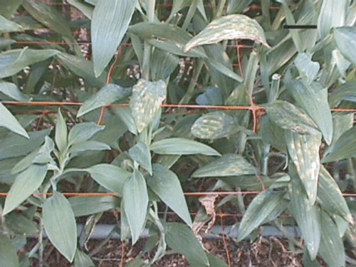
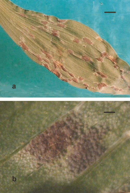
Symptoms consisted of spots, at first chlorotic with an oily appearance, then necrotic, irregular, scattered or confluent, and light brown in colour. They were located on the upper surfaces of the leaves, were enlarged and limited by the veins (Fig. 1). Conidia, conidiophores and stromata of a cercosporoid fungus developed on the lower surfaces of the leaves from the centre to the borders of the elongated spots (Fig. 2a, b). In severe cases, the petioles were also infected, the spots became confluent and the leaves died. The disease begins in the basal leaves and progresses to the upper ones (Fig. 1). It causes defoliation and reduces the aesthetic value of affected flowering branches, thereby reducing the market value of the cut flowers.
|
|
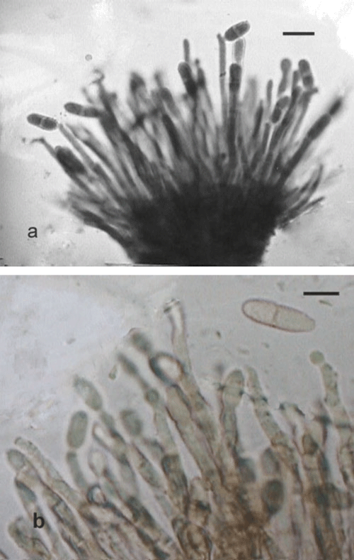
Colonies which developed on the leaves were hypophyllous, punctiform and confluent, olivaceous grey with new external colonies hyaline (Fig. 2b). Internal mycelia form stromata at first hyaline and then olivaceous grey. Conidiophores were grouped in dense fascicles, numerous to very numerous, sporodochial, erect, mostly straight, subcylindiric, unbranched, pale green-olivaceous, wall verrucose–subrugose, 105–150 μm (av. 117), three to six septa (Fig. 3a). Conidia were solitary, obpyriform, obclavate, 15–26 μm (av. 20.3) × 7–11 μm (av. 8.2), up three septa, but usually one septum, often constricted at the septa, pale olivaceous, rugose–verrucose, apex broadly rounded, base truncate, hylium somewhat thickened and darkened (Fig. 3a, b).
|
In an attempt to isolate the pathogen, pieces of affected leaves were surface-sterilised with 0.5% NaOCl for 1, 1.5 and 2 min and washed twice with sterile water. They were transferred to Petri dishes with potato dextrose agar (PDA), vegetables juice agar (V8A) and Alstroemeria leaf extract agar (AEA) (200 g Alstroemeria leaves, 20 g glucose, 20 g agar, 800 mL distilled water). In addition several colonies of the fungi emerging from the symptomatic leaves were transferred to slants of the same media. These methods were unsuccessful. Chambers and Fukenberg (1987) also failed to obtain isolates of Asperisporium caricae from infected pieces of sterilised leaf tissues on various tests media. They obtained satisfactory results only on 3 of 29 tested media using single conidia.
In the present study, isolates were obtained when leaf pieces were deposited on agar with their upper surfaces touching the media. Thus, new conidiophores and conidia which developed slowly on the leaf pieces after 20–25 days were transferred to slants of PDA and slowly developed dome shaped and compact colonies (3 mm in diameter at 35 days, 8 mm in diameter at 4 months).
Pathogenicity tests were conducted by the application of the inoculum suspension [105 propagules/mL with the addition of a tensiactive solution or water (controls)] with a brush, on potted plants of Alstroemeria hybrids cv. Rebeca. Inoculated plants were covered with plastic bags for 72 h and kept in a greenhouse at 17–23°C. Twenty-five days after inoculation, transluscent and chlorotic spots were observed on the leaves. Eight to ten days later, on the lower leaf surfaces, lesions turned olivaceous-grey as consequence of the formation of fungal colonies resembling natural infections. Leaves became chlorotic and died. The fungus re-isolated from these lesions was identical to that originally isolated from naturally infected plants.
Based on symptoms and cultural and morphological characteristics the fungus was identified as Asperisporium alstroemeriae (Allesch.) Maubl. This was confirmed by U. Braun, Martin-Luther-Universität Institut für Geobotanik und Botanischer Garten, Halle (Saale), Germany (material deposited at HAL 1752).
The wild Alstroemeria species, for example A. psittacina, could be a natural inoculum reservoir from where the pathogen is able to spread to commercial plantings. Preliminary observations in several greenhouses, where different cultivars of Alstroemeria hybrids were cropped, showed that all cultivars were susceptible to leaf spots. The only record found of this pathogen, named as Scolicotrichum alstroemeriae Allesch., was cited by Viégas on Alstroemeria spp. in South America (Viégas 1961). This is the first report of A. alstromeriae on commercial Alstroemeria spp. and on A. psittacina in Argentina. It appears to be also the first record on both hosts in the world.
Chambers KR, Fukenberg FHJ
(1987) Culture of Asperisporium caricae, the papaya black spot organism. Phytophylactica 19, 113.

Rollán MC,
Wolcan SM, Ronco L
(2005) First report of Uromyces alstroemeriae, causal agent of Alstroemeria rust in Argentina. Australasian Plant Pathology 34, 595–597.
| Crossref | GoogleScholarGoogle Scholar |