

First report of Rhizoctonia solani AG 4 HG-II attacking Gazania rigens plants in Brazil
D. D. Rosa A B , C. T. Ohto A , M. A. Basseto A , E. L. Furtado A and N. L. de Souza AA São Paulo State University, College of Agronomic Science, Department of Plant Production – Plant Health Protection Sector, PO Box 237, Botucatu, SP 18610-307, Brazil.
B Corresponding author. Email: ddrosa@fca.unesp.br
Australasian Plant Disease Notes 3(1) 1-2 https://doi.org/10.1071/DN08001
Submitted: 17 September 2007 Accepted: 10 January 2008 Published: 30 January 2008
Abstract
Leaf yellowing, wilting, petiole rot, root rot and death of plants were the symptoms observed in Gazania plants in gardens in São Paulo State, Brazil. Rhizoctonia solani AG-4 HGII was identified as the causal agent. This is the first record of Rhizoctonia solani AG 4 HG-II on Gazania rigens in Brazil.
Gazania rigens (Gazania) is an herbaceous plant native to South Africa, which belongs to the family Asteraceae. Gazania is widely used as an ornamental in gardens as it has excellent growth during all seasons of the year in Brazil. In January 2007, symptoms including leaf yellowing, wilting, petiole rot and root rot, leading to the death of plants, were observed in Gazania plants in gardens. The presence of white mycelia was observed in dead plants and necrotic regions. Fragments of necrotic regions were collected and disinfested in 70% alcohol for 30 s and 2% sodium hypochlorite for 30 s, washed in distilled water, and then plated onto PDA culture medium and incubated at 24°C in the dark for 24 h (Fenille et al. 2005). The isolated fungus showed hyphal ramification angles of ~90°, basal constriction, a septum next to the lateral hyphae, and other typical characteristics such as sclerotia, consistent with Rhizoctonia spp. (Sneh et al. 1991). This isolate was deposited in the São Paulo State University Mycology Database.
Twenty randomly selected cells per isolate were analysed, and the number of nuclei per cell was counted. Prior to examination, hyphae vegetative cells were stained with a DAPI technique (Kulik and Dery 1995) and the hyphae were stained with safranin-O 0.03% and KOH 3% solution and then observed under brightfield microscopy at 400× magnification, for the visualisation of the anastomosis reaction (Yamamoto and Uchida 1982). Hyphae were considered to be compatible when at least five points on each of four slides per isolate showed C2 and C3 type-reactions (Carling and Leiner 1990). Anastomosis was regarded as positive when hyphae between the Gazania isolate and AG tester made contact with each other and their walls fused, with subsequent plasmolysis of adjacent cells (Macnish et al. 1993). Tester isolates of R. solani and its subgroups (AG 1, AG 2, AG 3, AG 4, AG 5, AG 6, AG 7, AG 8, AG 9, AG 10, AG 11, AG 12, AG 13 and AG BI), obtained from researchers from different parts of the world, were paired with all isolates collected.
Colonies of cream-coloured mycelia grew close to the surface of potato dextrose agar (PDA) and the isolate formed mostly irregular whitish-brown sclerotia, 3.0 to 8.1 mm (av. 4.4 mm) in diameter inside the medium. The hyphae measured 7.1 to 7.8 µm (av. 7.4 µm) in diameter. We did not observe clamp connections. The Gazania isolate was multinucleate, with from 9 to 17 nuclei per cell, providing further support for the isolate being identified as belonging to the species Rhizoctonia solani. We observed the occurrence of a C2 anastomosis reaction between the Gazania isolate and the standard isolate for anastomosis group 4 HG-II, which allowed the Gazania isolate to be identified as belonging to the species Rhizoctonia solani AG 4 HG-II.
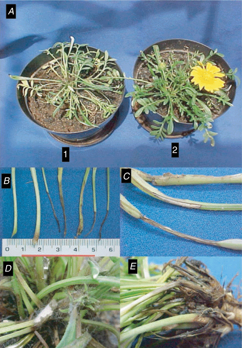
In a pathogenicity test, the Gazania isolate was grown in PDA culture medium for 5 days at 24°C in the dark. Three 5-mm-diameter disks were taken from the colonised culture medium and placed at the base of Gazania plants. Three Gazania plants were inoculated separately in a 1.5-L pot and another three non-inoculated plants were maintained as controls. After inoculation, both inoculated and non-inoculated plants were placed in a humid chamber at 22°C for 24 h, and later transferred to a greenhouse, in which the temperature was maintained at 25–27°C. Inoculated plants began to show yellowing, petiole necrosis, and root rot symptoms after 5 days (Fig. 1); all inoculated plants had died 15 days after inoculation. Sclerotia were not seen in the dead plants. No symptoms were observed in non-inoculated plants (Fig. 1). The pathogen was reisolated from fragments of necrotic regions of inoculated plants, and the presence of the fungus R. solani was demonstrated, thus confirming it as the causal agent. This is the first report of Rhizoctonia solani AG 4 HG-II attacking Gazania rigens in Brazil.
|
Acknowledgements
The authors wish to thank Dr J.B. Sinclair (University of Illinois, Urbana-Champaign, USA). Dr A. Ogoshi (Hokkaido University, Sapporo, Japan). Dr L. J. Herr (Ohio State University, Wooster, USA). Dr S. Naito (Tohoku National Agricultural Experiment Station, Morioka, Japan) and Dr M.A. Cubeta (Duke University, North Carolina, USA) for supplying the tester strains of R. solani.
Carling DE, Leiner RH
(1990) Virulence of isolates of Rhizoctonia solani AG-3 collected from potato plant organs and soil. Plant Disease 74, 901–903.
| Crossref | GoogleScholarGoogle Scholar |

Fenille RC,
Ciampi MB,
Souza NL,
Nakatani AK, Kuramae EE
(2005) Binucleate Rhizoctonia sp. AG G causing root rot in yacon (Smallanthus sonchifolius) in Brazil. Plant Pathology 54, 325–330.
| Crossref | GoogleScholarGoogle Scholar |
Kulik MM, Dery PD
(1995) Use of DAPI for anastomosis group typing of strains of the fungus Rhizoctonia solani. Biotechnique and Histochemistry 70, 95–98.
| Crossref | GoogleScholarGoogle Scholar |
Macnish GC,
Carling DE, Brainard KA
(1993) Characterization of Rhizoctonia solani AG-8 from bare patches by pectic isozyme (zymogram) and anastomosis techniques. Phytopathology 83, 922–927.
| Crossref | GoogleScholarGoogle Scholar |
Yamamoto DT, Uchida JY
(1982) Rapid nuclear staining of Rhizoctonia solani and related fungi with acridine orange and with safranin O. Mycologia 74, 145–149.
| Crossref | GoogleScholarGoogle Scholar |