

First report of bacterial necrosis of mango caused by Pseudomonas syringae pv. syringae in Australia
H. Golzar A C and E. J. Cother BA Department of Agriculture and Food Western Australia, Bentley Delivery Centre, WA 6983, Australia.
B NSW Department of Primary Industries, Agricultural Institute, Forest Road, Orange, NSW 2800, Australia.
C Corresponding author. Email: hgolzar@agric.wa.gov.au
Australasian Plant Disease Notes 3(1) 107-109 https://doi.org/10.1071/DN08043
Submitted: 10 June 2008 Accepted: 15 July 2008 Published: 23 July 2008
Abstract
A previously unreported bacterial disease was found on mango trees in the Gin-gin orchards north of Perth, Western Australia in 2007. The symptoms were necrosis of the stem tip, buds, flowers and leaf tissues. Based on biochemical tests, fatty acid analysis and pathogenicity tests, Pseudomonas syringae pv. syringae was identified as the causal agent of mango bacterial necrosis.
Pseudomonas syringae pv. syringae has been reported on a wide range of plant species including stone and pome fruits, citrus, bean, wheat, sugar and beet. The symptoms include dieback, bud and flower blast, spots and blisters on the fruit, stem cankers and leaf blight. Pseudomonas syringae pv. syringae has been reported as causing apical necrosis of mango (Mangifera indica L.) in southern Spain, Portugal and Israel. There, severe necrosis of buds and flower panicles have been shown to cause a significant decrease in fruit set but necrotic lesions have not been reported on fruit (Pinkas et al.1996; Cazorla et al.1998). Symptoms observed on mango trees in Perth orchards were similar to the previously reported symptoms. Pseudomonas syringae has been isolated from mango in northern NSW, and a similar bacterial disease is suspected to be affecting trees in the Bundaberg district of Queensland and Carnarvon in Western Australia (unpubl. data). However, there is no official record of mango bacterial necrosis caused by P. syringae pv. syringae in Australia.
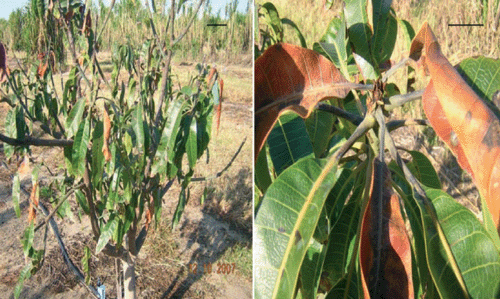
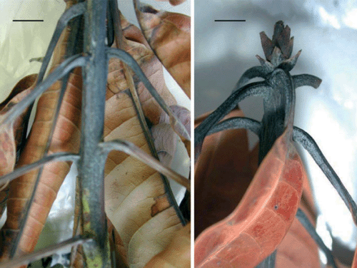
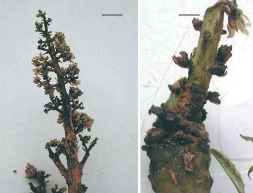
During winter and early spring in 2007, mango orchards in Gin-gin areas, north of Perth, Western Australia, were surveyed and samples with necrotic symptoms on the flowers and vegetative parts of the mango trees were randomly collected (Figs 1–3). Isolations were made from buds, panicles, leaf petioles, and necrotic lesions on the leaf and stem. Cream-coloured and fluorescent bacterial colonies were consistently isolated from the buds, stem tip, leaf petioles and leaf tissues adjacent to midribs and interveinal lesions. Isolates with positive hypersensitivity on tobacco (Nicotiana glutinosa) were used for further tests. The bacterial isolates from mango were identified based on biochemical tests (Lelliott and Stead 1987) and using the Biolog identification system based on the carbon utilisation microplate assay (Biolog MicroLog 4.0 System, Biolog Inc., Hayward, CA), fatty acid analysis (MIS-TSBA6) and pathogenicity tests (Lelliott and Stead 1987; Legard and Hunter 1990; Mazarei and Kerr 1990).
|
|
|
Isolates were Gram negative, fluorescent on King’s medium B, oxidase negative, catalase positive, potato soft rot negative, and arginine dihydrolase negative. Fatty acid methyl ester analysis characterised representative strains as P. syringae with similarity indices ranging from 0.77 to 0.97. These representative isolates were also tested using the Biolog system and were identified as P. syringae pv. syringae with a probability range of 95 to 100%. To confirm identification of the bacterial strains, pathogenicity tests with three mango isolates were performed on immature lemon, pear, zucchini fruit, young bean pods and tomato seedlings. Isolates were grown on sucrose peptone agar (SPA) for 24 h and then suspended in sterile water and diluted to a concentration of 106 CFU/mL. Fruits and bean pods were surface-sterilised with alcohol, washed with sterile water and inoculated by placing drops of an aqueous bacterial suspension on the surface and pricking through the drops using sterile needles. Controls were inoculated with sterile water. After inoculation, fruits and bean pods were incubated in the moist trays at 25°C for 7 days. Four-week-old healthy tomato plants were inoculated using the same bacterial inocula, controls and techniques. Plants were placed under mist for 48 h and then moved to the glasshouse at 22 ± 1°C. Disease symptoms were checked 6 days after inoculation.
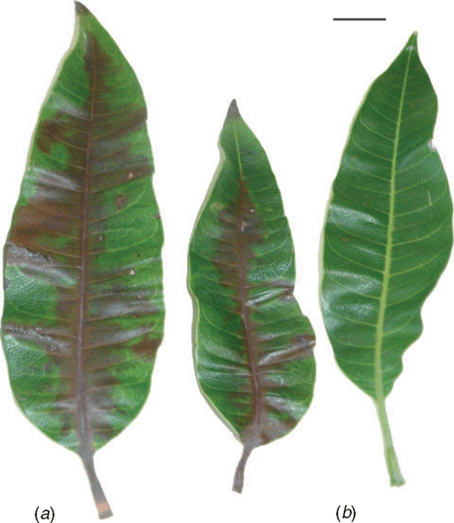
To test pathogenicity of the same P. syringae pv. syringae isolates on mangoes, young detached leaves were inoculated by placing drops of bacterial suspension (106 cfu/mL) on freshly wounded midribs. Controls were inoculated in the same way using sterile water. Inoculated leaves were incubated in moist trays at 25°C. Necrosis developed along the midribs and intervein tissues 7 days after inoculation (Fig. 4). The isolates also caused deep black, necrotic lesions on lemon fruits, dark sunken spots on pears and water soaked lesions on bean pods and zucchini fruit. Tomato leaves showed necrotic lesions 5 days after inoculation. Symptoms described above were similar to the symptoms previously reported on differential hosts in response to P. syringae pv. syringae strains (Lelliott and Stead 1987; Scheck et al. 1997; Cazorla et al. 1998). Koch’s Postulates were fulfilled and reisolated bacterial colonies were identified as P. syringae pv. syringae. Cultures of P. syringae pv. syringae have been deposited in the Herbarium DAR, Orange NSW as DAR 77789 and WA culture collection as WAC 13101.
|
To our knowledge, this is first report of P. syringae pv. syringae on mango in Australia.
Acknowledgements
The authors would like to thank Ms Dominie Wright for her help and Dr Jill Wilson for surveying mango orchards and providing mango samples. We acknowledge the technical assistance provided by Ms Paula Mather, Megan Jordan and Dorothy Noble; Dr Manisha Shankar for reviewing of the manuscript and the mango industry in Gin-gin for financial support.
Cazorla FM,
Tores JA,
Olalla L,
Perez-Garcia A,
Farre JM, de Vincente A
(1998) Bacterial apical necrosis of mango in southern Spain: a disease caused by Pseudomonas syringae pv. syringae. Phytopathology 88, 614–620.
| Crossref | GoogleScholarGoogle Scholar |

Legard DC, Hunter JE
(1990) Pathogenicity on bean of Pseudomonas syringae pv. syringae recovered from the phylloplane of weeds and from bean crop residue. Phytopathology 80, 938–942.
| Crossref | GoogleScholarGoogle Scholar |
Mazarei M, Kerr A
(1990) Distinguishing pathovars of Pseudomonas syringae on peas: nutritional, pathogenicity and serological tests. Plant Pathology 39, 278–285.
| Crossref | GoogleScholarGoogle Scholar |
Pinkas Y,
Maymon M, Smolewich Y
(1996) Bacterial black blight of mango. Alon Hanotea 50, 475.
Scheck HJ,
Canfield ML,
Pscheidt JW, Moore LW
(1997) Rapid evaluation of pathogenicity in Pseudomonas syringae pv. syringae with a lilac tissue culture bioassay and syringomycin DNA probes. Plant Disease 81, 905–910.
| Crossref | GoogleScholarGoogle Scholar |