

First report of Alternaria alternata causing leaf spot on Aloe barbadensis in India
A. Kamalakannan A , C. Gopalakrishnan A B , R. Renuka A , K. Kalpana A , D. Ladha Lakshmi A and V. Valluvaparidasan AA Department of Plant Pathology, Tamil Nadu Agricultural University, Coimbatore 641 003, Tamil Nadu, India.
B Corresponding author. Email: pc_gopal@yahoo.co.in
Australasian Plant Disease Notes 3(1) 110-111 https://doi.org/10.1071/DN08044
Submitted: 21 March 2008 Accepted: 24 July 2008 Published: 6 August 2008
Abstract
Aloe (Aloe barbadensis), an important medicinal plant grown in the state of Tamil Nadu, India has suffered heavy losses due to a leaf disease in 2006. The symptoms observed were small, circular to oval dark brown necrotic sunken spots located mostly on the leaf tip, with average diameter of 1.0 mm and reaching 3.0 mm. The pathogen was isolated and identified as Alternaria alternata and the pathogenicity was established. The conidiophores were branched, straight, golden brown in colour, measuring 15 µm long and 2–6 µm thick. The conidia were golden brown in colour, produced in long branched chains, obclavate in shape, with short conical flask. The literature indicates that this is the first report of a leaf spot disease of aloe in India.
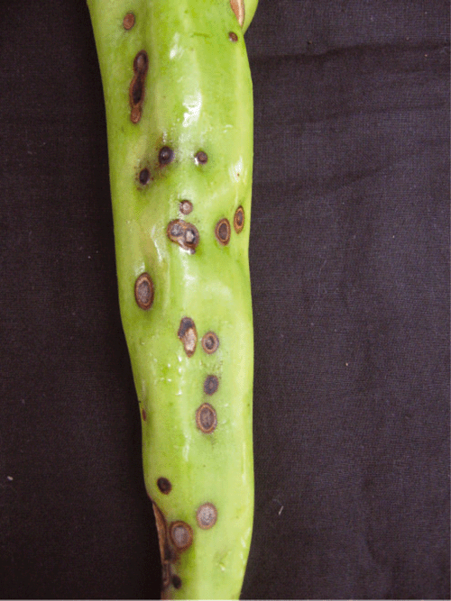
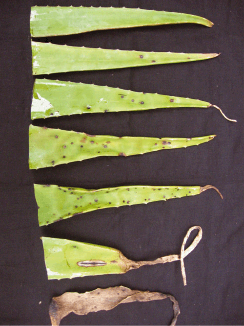
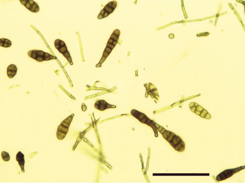
Aloe barbadensis (syn. Aloe vera) popularly known as Indian Aloe or Barbados Aloe is a perennial herb with fleshy leaves, which have great medicinal value. In June and July 2006, aloe grown in Coimbatore, Erode and Madurai districts of Tamil Nadu, India, suffered heavy losses due to a disease (Fig. 1). The symptoms observed were small circular to oval dark brown necrotic sunken spots located mostly on the leaf tip with an average diameter of 1.0 mm, reaching 3.0 mm (Fig. 2). The spots were characterised by having grey centres with brown margins. Black sporulation appeared in the centre of the spots. In a later stage of infection, the affected leaves dried from the tip downwards and lost the mucilaginous jelly (Fig. 3). The causal agent of this disease was successfully isolated on potato dextrose agar (PDA) from the diseased leaves. In total, 16 samples of infected leaves were collected from different parts of Tamil Nadu state and these infected leaves were used for isolations. The target pathogen was isolated from 11 samples. No other pathogenic fungus was isolated from the infected aloe leaves used in the test. After 4 to 5 days of incubation at 25 ± 2°C with a 12-h photoperiod, the fungus produced grey colonies with olive green peripheries. The conidiophores were branched, straight, golden brown in colour, measuring 15 µm long and 2–6 µm thick. The conidia were golden brown in colour and produced in long branched chains, obclavate in shape with short conical flask. The size of conidia varied from 22.75 to 63.70 µm in length and 13.65 to 18.20 µm in width with an average beak length of 7.73 µm. Conidia had two to three transverse septa and usually several longitudinal septa (Fig. 4).

|
|
|
|
Based on the symptoms, mycelial and conidial characters, the fungus was identified as Alternaria alternata (Ellis 1971). To confirm pathogenicity, a spore suspension (5 × 10⁴ conidia/mL) was sprayed onto pinpricked leaf pieces in Petri dishes lined with moist cotton. Detached pinpricked leaf pieces in Petri dishes lined with moist cotton and sprayed with sterile distilled water served as controls. The Petri dishes with leaf pieces were incubated at 25 ± 2°C under laboratory conditions. The pinpricked leaf pieces that were inoculated with spore suspension produced typical necrotic spots after 4 days of incubation. The fungus was consistently reisolated from infected leaf pieces onto PDA. In total, 10 leaf pieces were used in detached leaf assay studies and the pathogen was successfully isolated from all of them. In the present study, pathogenicity on leaf pieces without pinpricking was not investigated. However, pathogenicity was confirmed on pinpricked leaves grown in a glasshouse. Twenty plants were grown in pots and two leaves from each plant were randomly selected, pinpricked, sprayed with a spore suspension of the right concentration and incubated under glasshouse conditions. Pinpricked leaves sprayed with sterile distilled water served as controls. Typical symptoms were produced after 4–7 days on 32 leaves. Fifteen infected leaves were used to reisolate the pathogen. The pathogen was reisolated from all of them. In contrast, the control leaf did not show any symptoms.
Alternaria alternata has previously been reported as a leaf spot pathogen of tomato (Akhtar et al. 2004), stevia (Maiti et al. 2006) and pomegranate (Madhukar and Reddy 1976). To the best of our knowledge, this is the first report of leaf spot disease of aloe in India. A culture of A. alternata has been deposited in the culture collection of Department of Plant Pathology, Tamil Nadu Agricultural University, India.
Acknowledgements
The authors would like to thank P. Kalaiarasan and M. Sivakumar, Department of Nematology, TNAU, Coimbatore-3 for their excellent technical assistance in conducting research.
Akhtar KP,
Saleem MY,
Asghar M, Haq MA
(2004) New report of Alternaria alternata causing leaf blight of tomato in Pakistan. Plant Pathology 53, 816–816.
| Crossref | GoogleScholarGoogle Scholar |
[Verified 31 July 2008]