

Notes on Pseudomonas syringae pv. syringae bacterial necrosis of mango (Mangifera indica) in Australia
A. YoungCentre for Tropical Agriculture, Department of Primary Industries and Fisheries, 28 Peters Street, Mareeba, Qld 4880, Australia. Email: anthony.young@dpi.qld.gov.au
Australasian Plant Disease Notes 3(1) 138-140 https://doi.org/10.1071/DN08053
Submitted: 10 September 2008 Accepted: 24 September 2008 Published: 9 October 2008
Abstract
This paper provides current information on the epidemiology of the important mango disease, bacterial necrosis, caused by Pseudomonas syringae pv. syringae. It clarifies certain details contained within the first Australian report and provides further information on the symptoms and known epidemiology of the disease, in addition to potential management strategies.
History of mango bacterial necrosis in Australia
Mango bacterial necrosis was discovered by Dr Steve Akiew in the Bundaberg district of Queensland in September 1999, and in Byron Bay, New South Wales, in December of that year (Anon. 2000; S. Akiew, unpubl. data). While it affected all aerial parts of the plant, it was particularly apparent and damaging in the panicles, and so was then termed ‘panicle disease’ or ‘panicle blight’. Its main symptoms were blighting and necrosis of the panicle, resulting in reduced flowering and reduced-to-absent fruit set. A fluorescent pseudomonad was consistently isolated from infected panicles and was identified as Pseudomonas syringae using the LOPAT tests (Lelliot et al. 1966; S. Akiew, pers. comm.). The disease was then called ‘Pseudomonas flower blight’ and was included in the Queensland DPI&F Mango Disease Workshop Manual in 2001. The same disease has been identified in Israel, where it is known as ‘bacterial black blight’ (Pinkas et al. 1996), and in Spain and Portugal, where it is known as ‘bacterial apical necrosis’ (Cazorla et al. 1998, and references therein).
In late September 2007, the author was alerted to a destructive infection of mango near Casino, northern New South Wales, that was shown to be caused by P. syringae. The isolates were Gram negative, oxidase negative, potato soft-rot negative, catalase positive, fluorescent on King’s B medium (King et al. 1954) and motile via 1–2 polar flagella shown by silver impregnation (Blenden and Goldberg 1965). Suspecting it to be the same disease previously observed in the south-eastern growing districts and overseas, researchers from New South Wales, Queensland and Western Australia were contacted and photographs of the symptoms were distributed in order to determine whether or not the disease was present in other regions. It was soon established that the disease was also severe in the Gingin and Dandaragan areas of Western Australia.
In December 2007, a collaborative research proposal (MG08001, A. Young) was submitted to Horticulture Australia Limited (HAL) to better characterise the disease. Part of the summary was published in the Winter 2008 edition of the industry magazine ‘Mango Matters’ (Tyas 2008). However, researchers in Carnarvon, Western Australia, notified the author to an error in that the disease had not been identified in that region (A. Mackie, pers. comm.). The diagnosis of P. syringae was confirmed by fatty acid methyl ester (FAME) analysis, and an isolate was deposited in the Herbarium DAR, Orange, NSW as DAR 77787 (R. Cother, pers. comm.).
A recent paper by Golzar and Cother (2008) provides the first journal article of the presence of the disease in Australia. Their isolate was deposited in the Herbarium DAR, Orange, NSW as DAR 77789. Although of arguably trivial significance, the town of Gingin, Western Australia, is consistently misspelt throughout the article, which is here clarified to avoid confusion with the Queensland town of Gin Gin, where the disease has also been recorded on mango. A second point that requires clarification is that Carnarvon, Western Australia, is included in the distribution of the disease, albeit as unpublished data of unknown provenance. As discussed above, this was dismissed by Carnarvon mango researchers who first noticed the error in the HAL project proposal summary. It is important that this is rectified because it may confound future epidemiologists given the climatic conditions associated with the disease (see below), and it could potentially impact on future market access.
Symptoms and epidemiology
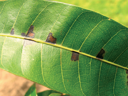
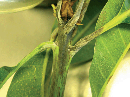
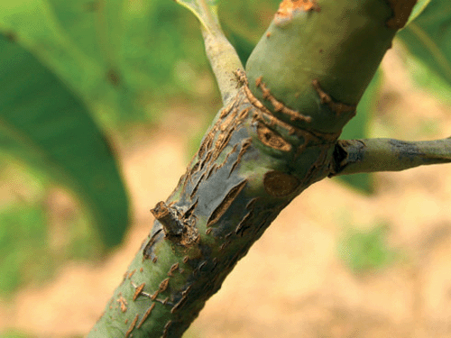
There are several characteristics that distinguish P. syringae bacterial necrosis from other mango diseases, but it is possible to confuse them. In Casino, New South Wales, the disease was originally diagnosed as anthracnose (Colletotrichum gloeosporioides) and bacterial black spot (Xanthomonas campestris pv. mangiferaeindicae), and so not surprisingly the plants did not respond to the standard treatments for these diseases, which included mancozeb, copper sprays and ultimately Amistar® (M. Coleman, pers. comm.). The infection does not appear to cause any blemish on fruits (when set), and is otherwise readily distinguished from anthracnose by the absence of fungal spores and having smaller, angular lesions that are at first delimited by the primary and secondary leaf veins. Young lesions are often observed in an alternating arrangement down either side of the leaf mid-rib (Fig. 1), but as they get older they may coalesce and cause complete leaf death. The apical meristem is often blighted (Fig. 2). There is evidence for cankering of the bark (Fig. 3), but the involvement of P. syringae has not yet been proven. Symptoms are distinct from bacterial black spot in that panicle, stem, petiole and leaf lesions are not clearly raised, do not have a chlorotic halo and do not produce obvious oozing or weeping. Microscopic examinations of transverse sections reveal only slight bacterial oozing.
|
|
|
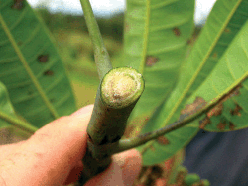
Bacterial necrosis appears to be systemic. In addition to the interveinal arrangement of foliar lesions, discoloured vasculature has been traced down petioles and infected stems (Fig. 4). This is thought to be epidemiologically significant because it may provide internal refuge for the bacteria during unfavourable conditions. While there is evidence of differing cultivar reactions (Pinkas et al. 1996; Anon. 2000; Cazorla et al. 2006), there has been no reported research to date on the impact on varieties grown in the Australian industry. There is anecdotal evidence that the cultivar Kensington Pride is particularly susceptible, but the disease has also been observed on Keitt, Calypso, Honey Gold, Nam Doc Mai and R2E2 varieties. It is not known whether any varieties grown in Australia are resistant.
|
There is a strong environmental correlation with symptom expression. Disease onset is associated with colder, wetter winters, and the occurrence of hail, rain and/or heavy dews has been linked to disease transmission (M. Coleman pers. comm., T. Campbell pers. comm., T. Cooke pers. comm., Cazorla et al. 1998). This is consistent with an observed peak in total bacterial populations in mango trees during colder months (Cazorla et al. 2006), and the ability of the bacterium to continue to grow at a much reduced rate in culture at 4°C. The ice nucleation properties of P. syringae pv. syringae have been implicated in exacerbating frost injury in other tree crops (Kennelly et al. 2007). The psychrophilic nature of the bacterium may explain the absence of disease symptoms from warmer mango-growing regions such as north Queensland, the Northern Territory and northern Western Australia. Winter rain may be critical to symptom expression, but it is possible that symptomless chronic infections are responsible for otherwise unaccounted losses of production. While wind and water spatter are presumed to be the major forms of transmission of P. syringae pv. syringae in mango, it is likely that flower-visiting animals such as birds, bats and insects may spread the infection between trees. There is no evidence that the bacterium can survive in crop debris, but it is possible that this may constitute another source of infection.
There is no information about the origin of bacterial necrosis. P. syringae pv. syringae has been reported from over 180 different monocotyledonous and dicotyledonous plants (Bradbury 1986), so it may have independently crossed into mango from several sympatric hosts. However, as the pathovar is potentially highly heterogeneous, it is also possible that the mango isolates represent distinct strains that have not yet been discriminated from other P. syringae pv. syringae isolates (Kennelly et al. 2007). There has been no published attempt at comparing the mango isolates from different regions, nor has it been established whether other P. syringae pv. syringae isolates can infect and cause disease in mango. The origin of inoculum sources needs to be ascertained in order to develop sustainable management strategies for mango bacterial necrosis.
Management
There are currently no known control measures for bacterial necrosis of mango. Despite previous work trialling various agricultural chemicals and plant defence activators, very little control has been achieved (Cazorla et al. 2006; A. Mackie, pers. comm.). Current stonefruit practice of copper sprays and heavy pruning may not be applicable to mango because of seasonal differences in flowering and fruiting cycle. Copper sprays targeted just before rainfall events have enjoyed some success in controlling P. syringae diseases of stonefruit, but these methods have not been attempted in mango. There is also the danger of copper toxicity if these applications occur over several years.
In the US, several bacterial diseases of fruit trees are controlled by either topical or injected applications of the antibiotic streptomycin. Although this practice is currently prohibited in Australia, it has the advantage of acting systemically (Crosse and Garrett 1958), and therefore, has the potential to be useful against P. syringae pv. syringae infections of mango. However, the development of resistance may preclude the long-term efficacy of these treatments (Young 1977; Sundin et al. 1994). An alternative is the use of a biological control agent against P. syringae pv. syringae. The common plant epiphytic bacterium Pantoea agglomerans has shown promise for the management of fire blight (Erwinia amylovora) of pome fruit (Wodzinski et al. 1994), and it is possible that biological control organisms will be discovered for bacterial necrosis of mango.
Any physical damage to the plant could possibly transmit the disease, so hygienic use of pruning instruments is necessary to prevent spread between plants. Care should be taken when moving machinery from known infected areas to uninfected areas, both within and between orchards. While copper treatments may not operate in a bactericidal manner (Cazorla et al. 2006), they may offer prophylactic treatment if applied just before and immediately after pruning. It is hoped that planned research and trial work will further improve our understanding of bacterial necrosis and lead to sustainable management of the disease.
Acknowledgements
The author would like to thank various collaborative researchers who, by being open to discussion, forthright with observations, and generous with their time, have made contributions to the knowledge base for this disease. Particularly Steve Akiew, Julie Harris, Alison Mackie, Neil Lantzke, Jill Wilson, Peter Trevorrow, Kathy Grice, Rowland Holmes, Tony Cooke, Bill Hatton, Rob Vennard, Peter Johnson, Ian Bally, Terry Campbell and Myke and Pamela Coleman.
Anon.
(2000) New mango disease. Mango Care 29, 12.

Blenden DC, Goldberg HS
(1965) Silver impregnation stain for Leptospira and flagella. Journal of Bacteriology 89, 899–900.
| PubMed |
Cazorla FM,
Tores JA,
Olalla L,
Perez-Garcia A,
Farre JM, de Vincente A
(1998) Bacterial apical necrosis of mango in southern Spain: a disease caused by Pseudomonas syringae pv. syringae. Phytopathology 88, 614–620.
| Crossref | GoogleScholarGoogle Scholar |
Cazorla FM,
Arrebola E,
Olea F,
Velasco L,
Hermoso JM,
Perez-Garcia A,
Tores JA,
Farre JM, de Vincente A
(2006) Field evaluation of treatments for the control of the bacterial apical necrosis of mango (Mangifera indica) caused by Pseudomonas syringae pv. syringae. European Journal of Plant Pathology 116, 279–288.
| Crossref | GoogleScholarGoogle Scholar |
Crosse JE, Garrett CME
(1958) Experiments on the movement of streptomycin in cherry trees. The Annals of Applied Biology 46, 310–320.
| Crossref | GoogleScholarGoogle Scholar |
Golzar H, Cother E
(2008) First report of bacterial necrosis of mango caused by Pseudomonas syringae pv. syringae in Australia. Australasian Plant Disease Notes 3, 107–109.
Kennelly MM,
Cazorla FM,
de Vincente A,
Ramos C, Sundin GW
(2007)
Pseudomonas syringae diseases of fruit trees: progress toward understanding and control. Plant Disease 91, 4–17.
| Crossref | GoogleScholarGoogle Scholar |
King EO,
Ward MK, Raney DE
(1954) Two simple media for the demonstration of pyocyanin and fluorescein. Journal of Laboratory and Clinical Medicine 44, 301–307.
| PubMed |
Lelliot RA,
Billing E, Hayward AC
(1966) A determinative scheme for the fluorescent plant pathogenic pseudomonads. Journal of Applied Bacteriology 29, 470–489.
| PubMed |
Pinkas Y,
Maymon M, Smolewich Y
(1996) Bacterial black blight of mango. Alon Hanotea 50, 475.
Sundin GW,
Demezas DH, Bender CL
(1994) Genetic and plasmid diversity within natural populations of Pseudomonas syringae with various exposures to copper and streptomycin bactericides. Applied and Environmental Microbiology 60, 4421–4431.
| PubMed |
Wodzinski R,
Umholtz TE,
Rundle JR, Beer SV
(1994) Mechanisms of inhibition of Erwinia amylovora by Erw. herbicola in vitro and in vivo. Journal of Applied Bacteriology 76, 22–29.
Young JM
(1977) Resistance to streptomycin in Pseudomonas syringae from apricot. New Zealand Journal of Agricultural Research 20, 249–251.