

First report of Colletotrichum acutatum and C. gloeosporioides causing leaf spots of olives (Olea europaea) in Australia
V. Sergeeva A B , R. Spooner-Hart A and N. G. Nair AA Centre for Plant and Food Science, University of Western Sydney, Locked Bag 1797, Penrith South, DC, NSW 1797, Australia.
B Corresponding author. Email: v.sergeeva@uws.edu.au
Australasian Plant Disease Notes 3(1) 143-144 https://doi.org/10.1071/DN08055
Submitted: 1 August 2008 Accepted: 15 October 2008 Published: 5 November 2008
Abstract
We report the occurrence of Colletotrichum acutatum and C. gloeosporioides on leaves of olives (Olea europaea) for the first time in Australia.
Anthracnose caused by Colletotrichum acutatum and C. gloeosporioides is a common and widespread fruit rot disease of olives (Olea europaea L.) in most olive growing regions in the world. The first record of C. gloeosporioides on olive fruit in Australia was from Wagga Wagga, NSW, in 1969 (DAR 17788) and that of C. acutatum on fruit at both Roseberry, NSW (DAR 64937) and Parramatta, NSW (DAR 65049) in 1989. We report the occurrence of anthracnose caused by C. acutatum and C. gloeosporioides on leaves of olives (Olea europaea) for the first time in Australia.
Anthracnose has been previously recorded on olive leaves in Mediterranean countries such as Portugal, Spain and Italy (Tjamos et al. 1993; Talhinhas et al. 2005). Olive anthracnose caused by Colletotrichum spp. is becoming an important disease of fruits causing major losses in yield of olives and the quality of olive oil (Iannota et al. 1999; Talhinhas et al. 2005).
Leaf spot disease caused by C. acutatum J.H. Simmonds and C. gloeosporioides (Penz.) Penz. & Sacc. was observed in Australia in July 2007 on 8–10 years old olive trees of var. Barnea and var. Manzanillo with a case history of anthracnose disease. The trees were located in two olive groves in New South Wales at Laguna (32.995°S, 151.134°E), and Menangle (34.123°S, 150.743°E). The isolates of C. acutatum from var. Barnea and var. Manzanillo were deposited in the New South Wales Plant Pathology Herbarium as DAR 78445 and DAR 78877, respectively, and those of C. gloeosporioides from var. Manzanillo as DAR 78878.
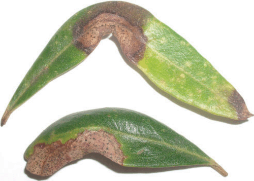
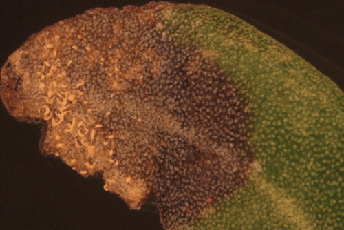
Anthracnose infection starts as small spots on the upper surface of leaves which can enlarge to form extensive dead areas. Brown spots carrying heavily sporulating colonies of C. acutatum (Fig. 1) and C. gloeosporioides (Fig. 2) were observed. The infected spots were observed mostly at the edges of the leaves but also occurred near the midrib. The spots were light to dark brown in colour and the necrotic areas had irregular shapes and were 1–3 cm in size. The acervuli on infected leaves produced salmon coloured spore masses under high relative humidity. Symptoms associated with leaf infection by C. gloeosporioides were similar to those caused by C. acutatum. Conidiomata are visible on leaves as small black dots.
|
|
Spores were observed after incubation of infected leaves for 2–3 days in a humid chamber at 25°C. Colletotrichum acutatum produced orange to pink coloured colonies with whitish aerial mycelium on potato dextrose agar (PDA). Colletotrichum gloeosporioides produced grey colonies with whitish aerial mycelium on PDA.
Leaves showing the symptoms were surface sterilised with 1% sodium hypochlorite for 2 min followed by washing three times in sterile distilled water. The fungal isolate was cultured on PDA and grown at 25°C. Pathogenicity tests were carried out by using conidial suspensions of C. acutatum and C. gloeosporioides. Individual, uninfected leaves on 5-year-old trees of variety Manzanillo growing in glasshouse at 25°C were used for pathogenicity tests. Suspensions of conidia (4 × 10⁶/mL) were prepared in sterile distilled water using 14-day-old cultures of the fungi and were used for spraying the leaves. The leaves on the trees were sprayed with sterile water served as controls. Both the inoculated and control leaves were covered with plastic bags and left for three days. Symptoms recorded after 7–9 days were similar to those initially observed on the trees. No symptoms developed on the leaves sprayed with sterile water. Colletotrichum acutatum and C. gloeosporioides were consistently reisolated from surface sterilised leaves of inoculated plants indicating internal infections of these leaves.
The incidence of naturally occurring infection on leaves by C. acutatum and C. gloeosporioides was relatively low in the different olive varieties and locations of olive groves. Data on incidence of leaf infection of 50 leaves each from 20 trees of var. Barnea at Menangle, showed that natural infection was 1–2% of leaves per tree. Heavy infections of anthracnose cause massive defoliation (Graniti et al. 1993). The leaf infections at Laguna and Menangle were observed during July, August, November, January and April when the leaves were collected for examination. However, it is very likely that the pathogens were present during the whole season. The lengthy presence of C. acutatum and C. gloeosporioides can be a continuing source of flower and fruit infection (Sergeeva et al. 2008). Infected leaves were considered as inoculum sources for fruit anthracnose (Cacciola et al. 1996). This is the first report of C. acutatum and C. gloeosporioides causing leaf spot in olive in Australia.
This work was part of a project funded by Horticulture Australia Limited, NuFarm Australia and Boundary Bend Management.
Cacciola SO,
Agosteo GE,
Pane A, Magnano di San Lio G
(1996) Osservazioni sull’epidemiologia dell’antracnosi dell’olivo in Calabria. Informatore Fitopatologico 46, 27–32.

Graniti A,
Frisullo S,
Pennisi AM, Magnano di San Lio G
(1993) Infection of Glomerella cingulata on olive in Italy. OEPP/EPPO Bulletin 23, 457–465.
Iannota N,
Perri E,
Serianni R, Tocci C
(1999) Influence of Colletotrichum gloeosporioides (Penzig) and Camarosporium dalmatica (Thum) attacks on olive quality. Acta Horticulturae 474, 573–576.
Sergeeva V,
Nair NG, Spooner-Hart R
(2008) First report of flower infection in olives (Olea europaea) by Colletotrichum acutatum and C. gloeosporioides causing anthracnose disease. Australasian Plant Disease Notes 3, 81–82.
Talhinhas P,
Sreenivasaprasad S,
Neves-Martin J, Oliveira H
(2005) Molecular and phenotypic analyses reveal association of diverse Colletotrichum acutatum groups and a low level of C. gloeosporioides with olive anthracnose. Applied and Environmental Microbiology 71, 2987–2998.
| Crossref | GoogleScholarGoogle Scholar | PubMed |
Tjamos EC,
Graniti A,
Smith IM, Lamberti F
(1993) Conference on olive diseases. EPPO Bulletin 23, 365–550.
| Crossref |