First report of Pseudomonas viridiflava in basil seedlings and plants in soilless crop in Italy
A. Minuto A E , G. Minuto A , P. Martini B , M. Odasso B , E. Biondi C , S. Mucini C and M. Scortichini DA Ce.R.S.A.A., Albenga (SV), Regione Rollo, 98 Albenga, SV I-17031, Italy.
B I.R.F., Sanremo (IM), Via Carducci, 12 Sanremo, IM I-18038, Italy.
C Facoltà di Agraria (DiSTA), Università di Bologna, Viale G. Fanin 40, Bologna I-40127, Italy.
D C.R.A., Centro di Ricerca per la Frutticoltura, Via di Fioranello 52, Rome I-00134, Italy.
E Corresponding author. Email: minuto.andrea@tiscali.it
Australasian Plant Disease Notes 3(1) 165-165 https://doi.org/10.1071/DN08064
Submitted: 5 November 2008 Accepted: 11 November 2008 Published: 27 November 2008
Abstract
Pseudomonas viridiflava is identified for the first time as a pathogen of soilless sweet basil seedlings in Italy.
Sweet basil (Ocimum basilicum L.) cv. Genovese showing necrosis of cotyledons, leaves and stems was repeatedly and consistently observed in Northern Italy (Liguria region) in spring 2007 and winter 2007–08 where around 80 ha are grown.
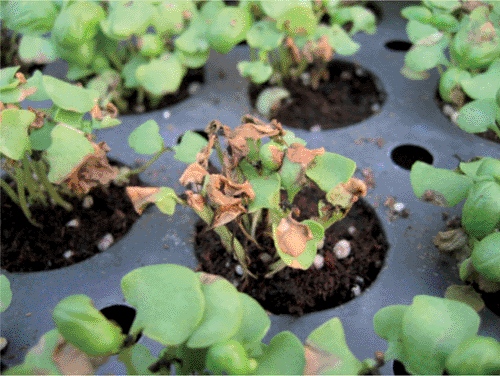
Symptomatic sweet basil plants were grown in trays and pots in a protected open ebb and flow system. Initial symptoms developed 10–14 days after sowing trays or after the emergence of the sixth to seventh leaf on potted plants. Symptoms consisted of dark grey, water-soaked, circular to irregular lesions of 2–3 mm, mainly on the leaf and cotyledon margins. Lesions eventually coalesced into brown-to-black areas and progressed to petioles and stems. Cotyledons abscised suddenly, and thereafter, plants withered and died within a few days (Fig. 1). More than 90% of plants exhibited symptoms.

|
Sections of symptomatic tissue from affected plants were surface disinfested in 0.5% NaOCl, washed in sterile deionised water, macerated in nutrient yeast dextrose broth, streaked onto nutrient yeast dextrose agar (NYDA) and incubated at 22 ± 1°C for 48 h. Pale yellow colonies were isolated on NYDA and transferred on King’s B medium (King et al. 1954) and subsequently eight fluorescent single cell colonies were purified. All isolates induced the hypersensitive reaction on tobacco leaves after 24–36 h. They were levan, oxidase, arginine dehydrolase-negative and showed pectolytic activity on potato tuber slices. Their repetitive-PCR pattern performed using BOX and ERIC primer sets were similar (homology: 92%) to strain NCPPB 635 of Pseudomonas viridiflava.
The pathogenicity of eight isolates was tested twice by spraying 15-day-old healthy basil plants with a bacterial suspension (106 cfu/mL). Two groups of 200 plants were respectively inoculated and sprayed with sterile nutrient broth as a control. Plants were kept at 25−28°C and 80–100% RH. Twenty-four to 36 h after inoculation, brown-to-black lesions were observed only on inoculated plants. Reisolations were made and resulting bacterial strains were identified as P. viridiflava.
The same bacterium has been reported in the United States (Little et al. 1994) and in Argentina (Alippi et al. 1999) in soil cultivation. A previous report described the rare presence of P. viridiflava in Sardinia, Italy, in soil (Franceschini et al. 1996), while to our knowledge, this is the first report of heavy infections of P. viridiflava in basil grown in hydroponic conditions in Liguria where sweet basil is now recognised as a Protected Designation of Origin crop by the European Union.
Alippi AM,
Wolcan S, Dal Bó E
(1999) First Report of bacterial leaf spot of basil caused by Pseudomonas viridiflava in Argentina. Plant Disease 83, 876.
| Crossref | GoogleScholarGoogle Scholar |

King EO,
Ward MK, Raney D
(1954) Two simple media for demonstration of pyocyanin and fluorescin. Journal of Laboratory and Clinical Medicine 44, 301–307.
|
CAS |
PubMed |

Little EL,
Gilbertson RL, Koike ST
(1994) First report of Pseudomonas viridiflava causing a leaf necrosis on basil. Plant Disease 78, 831.


