

Skin lesion suspicious of melanoma: time to one-step removal
Antonio Tejera-Vaquerizo 1 4 , Francisco Russo 2 , Gonzalo Nieto-González 31 Dermatology Department, Instituto Dermatológico GlobalDerm, Palma del Río, Córdoba, Spain.
2 Dermatology Department, Hospital Punta de Europa, Algeciras, Cádiz, Spain.
3 Dermatology Department, Hospital Santos Reyes, Aranda del Duero, Burgos, Spain.
4 Correponding author. Email: antoniotejera@aedv.es
Journal of Primary Health Care 11(2) 87-88 https://doi.org/10.1071/HC19044
Published: 18 July 2019
Journal Compilation © Royal New Zealand College of General Practitioners 2019.
This is an open access article licensed under a Creative Commons Attribution-NonCommercial-NoDerivatives 4.0 International License.
We read with interest the recent study by Brian and Jameson1 on compliance with clearance margins recommended by the Clinical Practice Guidelines for the Management of Melanoma in Australia and New Zealand. These guidelines recommend a 2 mm horizontal margin for cases of suspected melanoma treated with excision biopsy.2 The recommendation of similar margins in many countries is beginning to stir debate on this topic.3,4
The 2 mm recommendation is a low-grade recommendation (C) supported by an even lower level of evidence (IV).2 The main reason for such a small margin is not to interfere with lymphatic drainage as sentinel lymph node (SLN) biopsy may be indicated. However, SLN biopsy does not improve melanoma-specific survival,5 and neither does complete lymph node dissection following a positive biopsy, highlighting the need for reassessment of how primary melanomas should be managed.
One option that merits evaluation is one-step surgery for suspected in situ or thin melanomas as these account for a large proportion of incident cases of primary tumours.6 Although no tools are yet available for confirming a diagnosis of melanoma before excision, dermoscopy is very useful for distinguishing between nevus and melanoma in situ.7 The distinction between in situ and invasive melanoma is somewhat less clear.8
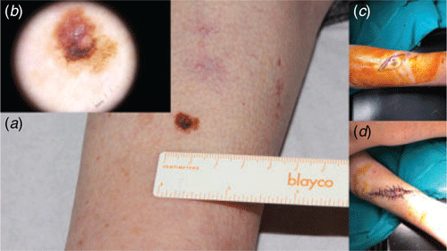
We propose performing one-step excision with a 10-mm horizontal clearance margin (Fig. 1) in patients with lesions that are strongly suspected to be melanoma located in suitable anatomic sites. This approach would avoid the need for re-excision in the case of thin melanomas (≤ 1 mm) or in situ melanomas. Its limitation is that the margin taken in the case of an in situ melanoma would be 5 mm larger than that recommended by clinical practice guidelines, but we believe that the aesthetic outcome is acceptable.
|
This one-step approach would reduce costs and could even have a favourable psychological impact, as some patients scheduled for re-excision believe that their prognosis has worsened.9
Conflicts of interest
The authors declare no conflicts of interest.
Funding
This research did not receive any specific funding.
References
[1] Brian T, Jameson MB. Skin lesions suspicious for melanoma: New Zealand excision margin guidelines in practice. J Prim Health Care 2018; 10 210–4.| Skin lesions suspicious for melanoma: New Zealand excision margin guidelines in practice.Crossref | GoogleScholarGoogle Scholar | 31039935PubMed |
[2] Aitken J, Barbour A, Burmeister B, et al. Clinical practice guidelines for the management of melanoma in Australia and New Zealand. Management 2008;
[3] National Comprehensive Cancer Network. Melanoma (Version 1.2019). [Cited 2019 January 15] Available from: https://www.nccn.org/professionals/physician_gls/PDF/melanoma.pdf.
[4] Russo F. Current evidence of one-step surgical treatment in cutaneous melanoma. Actas Dermosifiliogr 2019; In press
| 30982570PubMed |
[5] Morton DL, Thompson JF, Cochran AJ, et al. Final Trial Report of Sentinel-Node Biopsy versus Nodal Observation in Melanoma. N Engl J Med 2014; 370 599–609.
| Final Trial Report of Sentinel-Node Biopsy versus Nodal Observation in Melanoma.Crossref | GoogleScholarGoogle Scholar | 24521106PubMed |
[6] Elwood JM, Kim SJH, Ip KHK, et al. In situ and invasive melanoma in a high-risk, New Zealand, population: a population-based study. Australas J Dermatol 2019; 60 38–44.
| In situ and invasive melanoma in a high-risk, New Zealand, population: a population-based study.Crossref | GoogleScholarGoogle Scholar | 30051460PubMed |
[7] Lallas A, Longo C, Manfredini M, et al. Accuracy of dermoscopic criteria for the diagnosis of melanoma in situ. JAMA Dermatol 2018; 154 414–9.
| 29466542PubMed |
[8] da Silva VPM, Ikino JK, Sens MM, et al. Dermoscopic features of thin melanomas: a comparative study of melanoma in situ and invasive melanomas smaller than or equal to 1 mm. An Bras Dermatol 2013; 88 712–7.
| Dermoscopic features of thin melanomas: a comparative study of melanoma in situ and invasive melanomas smaller than or equal to 1 mm.Crossref | GoogleScholarGoogle Scholar |
[9] Kneier AW. The psychological challenges facing melanoma patients. Surg Clin North Am 1996; 76 1413–21.
| The psychological challenges facing melanoma patients.Crossref | GoogleScholarGoogle Scholar | 8977559PubMed |