

Diagnosis of internal carotid artery stenosis in a patient referred to a physiotherapist for dizziness
Ryan Boggs 1 3 , Michael Ross 1 , Michael Tall 21 Department of Physical Therapy, Daemen College, Amherst, NY, USA.
2 Department of Radiology, University of Texas Health Science Center at San Antonio, San Antonio, TX, USA.
3 Corresponding author. Email: rboggs1@daemen.edu
Journal of Primary Health Care 11(4) 373-379 https://doi.org/10.1071/HC19047
Published: 18 December 2019
Journal Compilation © Royal New Zealand College of General Practitioners 2019 This is an open access article licensed under a Creative Commons Attribution-NonCommercial-NoDerivatives 4.0 International License
Abstract
PURPOSE: The purpose of this report is to describe the diagnostic focus of the clinical decision-making process for a patient referred to a physiotherapist for treatment of persistent dizziness, who was subsequently diagnosed with severe stenosis of the internal carotid arteries.
CASE DESCRIPTION: The patient was a 79-year-old man who was referred to a physiotherapist by his primary care physician for the treatment of persistent intermittent dizziness. The patient’s dizziness began 6 months prior insidiously; it was worsening over time and now interfered with activities of daily living. The patient denied cervical pain or headaches, numbness or tingling in his extremities, difficulty maintaining balance with walking, unsteadiness, muscle weakness, dysphagia, drop attacks, diplopia or dysarthria. At the physiotherapist’s initial evaluation, cervical range of motion was moderately restricted in all motions and his dizziness was elicited with changes in head position. The patient’s neurological examination was unremarkable. Due to positional complaints of dizziness, a Dix–Hallpike test was used to screen for benign paroxysmal positional vertigo, which was positive for symptoms reproduction; however, no nystagmus was noted. The patient also became diaphoretic and exhibited significant discoloration of his face during the test.
OUTCOMES: Due to concern over vascular compromise, carotid duplex ultrasonography and magnetic resonance angiography were completed and revealed near complete occlusion of the left internal carotid artery at its origin. The patient subsequently underwent a left internal carotid endarterectomy with resolution of symptoms and a return to all activities of daily living.
DISCUSSION: Carotid artery stenosis, although frequently asymptomatic until severe, may manifest as complaints of dizziness that mimic peripheral vestibular dysfunction. Appropriate and prudent screening and referral is necessary if clinical symptoms suggestive of vascular compromise are present.
KEYwords: Dizziness; carotid artery stenosis; medical screening
Introduction
Dizziness is the chief presenting symptom in ~5% of primary care visits.1,2 Diagnosing the cause of dizziness can be difficult because symptoms are often non-specific and the differential diagnosis is broad (Table 1).3 One of the more common causes of dizziness and balance disorders in the elderly is benign paroxysmal positional vertigo (BPPV), which is caused by displacement of the otoconia from the otolith organ,4,5 mainly from the utricle, and the migration of this matter into one or more of the semicircular canals.6 The diagnosis of BPPV is clinical and done mainly through manoeuvres that determine which canal is involved and what type of BPPV is present. For example, the Dix–Hallpike test (Figure 1) evaluates the anterior and posterior canals and is considered the gold standard for the diagnosis of BPPV.7,8 If this test is positive, the person evaluated presents with vertigo and nystagmus.9 Patients with BPPV are commonly referred for vestibular rehabilitation, which is an effective, reliable and non-invasive therapeutic approach. Treatment success depends on identifying and specifying the type of BPPV and canal involved.10

|

|
It is important to differentiate dizziness requiring non-invasive symptom management and vestibular rehabilitation from conditions requiring further diagnostic work-up for serious, yet treatable, causes. Some consider dizziness the most difficult symptom to diagnose,11 and there is growing evidence that misdiagnosis of patients seen in an emergency department with dizziness is common.12,13 One example of a condition that may present as dizziness and mimic peripheral vestibular dysfunction, although frequently asymptomatic until severe, is carotid artery stenosis.14–16 The purpose of this report is to describe the diagnostic focus of the clinical decision-making process for a patient who was referred to a physiotherapist for treatment of persistent dizziness and subsequently diagnosed with severe stenosis of the internal carotid arteries.
Case report
The patient was a 79-year-old man referred to an outpatient physiotherapy practice by his primary care physician for the treatment of persistent intermittent dizziness that was thought to be related to BPPV and slowly worsening over time. The physiotherapist who evaluated the patient was a certified vestibular rehabilitation specialist. The patient’s chief complaint of dizziness began 6 months prior, insidiously after getting out of bed. He reported that he felt so dizzy and nauseous after his initial bout of dizziness that he stayed in bed for the next 3 days. His dizziness now interfered with activities of daily living. The patient denied cervical pain or headaches, numbness or tingling in his extremities, difficulty maintaining balance with walking, unsteadiness, muscle weakness, dysphagia, drop attacks, diplopia or dysarthria. The patient’s past medical history included hypertension and depression, for which he was taking metoprolol, 81 mg aspirin and mirtazapine. No diagnostic imaging was performed before the physiotherapist’s evaluation.
At the time of the physiotherapist’s initial evaluation, the patient had no complaints of dizziness at rest, his blood pressure was 126/62 mmHg, and he was independent with ambulation. Cervical range of motion was moderately restricted in all motions and his dizziness was elicited with changes in head position, especially cervical rotation in standing and supine. The patient’s neurological examination was unremarkable.
Due to positional complaints of dizziness, a Dix–Hallpike test (Figure 1) was used to screen for BPPV.7,8 Testing was positive for symptom reproduction with the head turned towards the right, which was the side tested first,17 but no nystagmus was noted. The patient also became diaphoretic and exhibited significant discoloration of his face during the test. The patient was returned to the sitting position and was closely monitored while his symptoms returned to baseline. Based on his subjective complaints and the physical examination findings, a primary diagnosis of BPPV was considered and treatment was initiated. The Dix–Hallpike test with the head turned towards the left was not performed.
After explaining the results of the Dix–Hallpike test, the patient and his daughter, who was present, gave consent for the physiotherapist to perform the Epley manoeuvre, which is a canalith repositioning technique and an effective treatment for BPPV.18 During the Epley manoeuvre, the patient reported symptoms of dizziness that dissipated after 30 s, nausea, and again became diaphoretic. He also reported a decrease in dizziness when returned upright from the Epley manoeuvre. The patient and his daughter were both educated by the physiotherapist about BPPV. He was asked to follow up with the physiotherapist 2 days later for further evaluation and re-assessment of his symptoms, but to contact the physiotherapist with any questions or concerns that may arise earlier.

|
On follow-up 2 days later, the patient reported that he experienced an episode of severe dizziness one day prior that eventually resolved with rest. Additionally, this episode of dizziness was accompanied with a reported loss of vision and syncope (Figure 2).
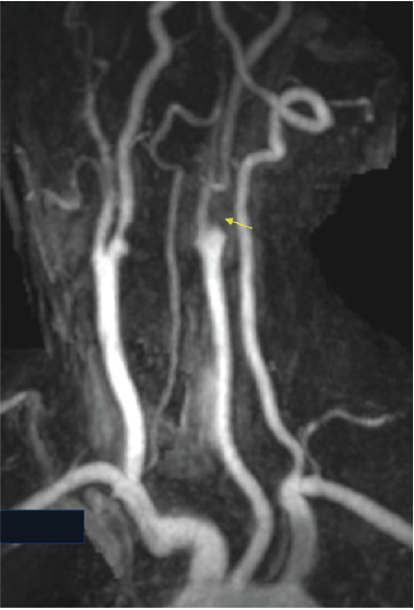
Given these new symptoms and concern over vascular compromise, the patient was immediately referred back to his primary care physician. Subsequent carotid duplex ultrasonography demonstrated stenosis of both the proximal left and right internal carotid arteries, with stenosis in the range of 50–69% bilaterally and slightly worse on the left (Figure 3).19 Magnetic resonance angiography revealed near complete occlusion of the left internal carotid artery at its origin (Figure 4). On the right side, there was narrowing of the right internal carotid artery correlating to a stenosis of 80%. The vertebral arteries were unremarkable bilaterally. The patient subsequently underwent a left internal carotid endarterectomy with resolution of symptoms and a return to all activities of daily living.

|
|
Discussion
The patient described in this report was referred to a physiotherapist by his primary care physician for treatment of persistent intermittent dizziness that was thought to be related to BPPV. By the time he was seen by the physiotherapist, his symptoms had been slowly worsening over the course of the previous 6 months after an initial onset of severe dizziness and nausea that occurred after getting out of bed (Figure 2). Although BBPV is a common cause of dizziness, the duration of symptoms may vary and can potentially persist for days, weeks or months or become recurrent over the course of many years.3
Complaints of dizziness may be a sign of more serious underlying vascular pathology, including a possible transient ischaemic attack or stroke.20 The typical clinical presentation of BPPV is a short duration of intense spinning vertigo when a patient lies down, turns the head to the side, looks up or down, or transitions upright from a supine position.7,8 This can be easily screened for with a Dix–Hallpike test and treated with a canalith repositioning technique (e.g. Epley manoeuvre) if the Dix–Hallpike test is positive for symptom reproduction. Autonomic symptoms such as pallor and diaphoresis commonly accompany peripheral vestibular disorders and may be present during testing.7 However, when these symptoms are associated with changes in visual acuity or feelings of syncope, temporary impaired blood flow to the brain should be considered as the root cause of symptoms and appropriate medical evaluation is indicated.
For the patient described in this report, diaphoresis and discoloration of his face were noted during the Dix–Hallpike test, but nystagmus was not noted. On follow up two days later, the patient reported he experienced an episode of severe dizziness the previous day that was accompanied with loss of vision and syncope. Given these new symptoms and concern over vascular compromise, the patient was immediately referred to his primary care physician for further medical evaluation.
Carotid duplex ultrasonography is the standard of care for the initial diagnosis of carotid artery bifurcation disease. As it is non-invasive and readily available, it is commonly the first diagnostic imaging modality used to screen patients with concerns for carotid artery stenosis. Carotid duplex ultrasonography has a sensitivity of 98% and specificity of 88% for the detection of >50% stenosis of the internal carotid artery. It has a sensitivity of 94% and specificity of 90% for detecting >70% stenosis of the internal carotid artery.21
If a patient’s carotid duplex ultrasonography reveals >50% stenosis of the internal carotid artery and the patient is being considered for invasive management strategies, magnetic resonance angiography is usually performed to allow for improved evaluation of the stenosis.22 Invasive treatment is considered for patients who are asymptomatic with stenosis >60%23,24 and patients who are symptomatic with stenosis >50%.25 The patient described in this report was initially evaluated with carotid duplex ultrasonography that demonstrated stenosis of both the proximal left and right internal carotid arteries, with stenosis in the range of 50–69% bilaterally and slightly worse on the left (Figure 3).19 These findings were subsequently confirmed with magnetic resonance angiography, which revealed near complete occlusion of the left internal carotid artery at its origin (Figure 4).
While near complete occlusion of the internal carotid artery is relatively rare with a prevalence of <10% among patients with significant carotid artery stenosis, it is an important cause of transient ischaemic attacks and stroke.26 Furthermore, patients undergoing evaluation for acute stroke should have carotid imaging due to the large proportion of embolic strokes (20–25%) that originate from the carotid artery.22 While carotid artery disease can be managed medically, surgically and with minimal invasive interventional procedures, carotid endarterectomy has become the standard treatment for patients with severe carotid artery stenosis.27 Previous studies examining the efficacy of carotid endarterectomy in stroke prevention for both symptomatic and asymptomatic carotid artery stenosis have shown promising short- and long-term outcomes for this procedure.28–32
When the patient described in this report was evaluated with the Dix–Hallpike test, his primary symptom of dizziness was reproduced and he became diaphoretic and exhibited significant discoloration of his face during the test. However, no nystagmus was noted. Alvarenga et al.33 recommend treatment of BPPV without nystagmus may be carried out based on the typical history of BPPV and signs and symptoms found during the history and physical examination (ie dizziness, nausea, diaphoresis). They further report that symptom remission among patients with BPPV without nystagmus suggests the need for BPPV treatment. Therefore, even though no nystagmus was noted with the Dix–Hallpike test, this patient was treated with the Epley manoeuvre because of the onset of his dizziness and diaphoresis with testing.18 However, the lack of improvement and the development of new concerning symptoms (loss of vision and syncope) lessened the likelihood that the patient’s symptoms were related to BPPV. Immediate medical referral was then initiated due to concern over vascular compromise.
The patient described in this report was referred to a physiotherapist by his primary care physician for the treatment of persistent intermittent dizziness that was thought to be related to BPPV. The patient’s evaluation, however, was not consistent with BPPV and he subsequently developed symptoms that raised concern of a transient ischaemic attack. These signs and symptoms of cardiovascular disease, together with his known history of cardiovascular risk factors (age, history of cardiovascular disease, taking anti-hypertensive medication), put him at high risk for a cardiovascular event and also made him a likely candidate for vascular pathology. Carotid artery disease should be considered as a potential underlying pathology in patients with such a profile.
Conclusion
Carotid artery stenosis, although frequently asymptomatic until severe, may manifest as complaints of dizziness that mimic peripheral vestibular dysfunction. Appropriate screening and referral are necessary if clinical symptoms suggesting vascular compromise are present.
Competing interests
The authors declare no competing interests.
References
[1] Sloane PD. Dizziness in primary care. J Fam Pract. 1989; 29 33–8.| 2738548PubMed |
[2] Kroenke K, Jackson JL. Outcome in general medical patients presenting with common symptoms: a prospective study with a 2-week and a 3-month follow-up. Fam Pract. 1998; 15 398–403.
| Outcome in general medical patients presenting with common symptoms: a prospective study with a 2-week and a 3-month follow-up.Crossref | GoogleScholarGoogle Scholar | 9848423PubMed |
[3] Post RE, Dickerson LM. Dizziness: a diagnostic approach. Am Fam Physician. 2010; 82 361–8.
| 20704166PubMed |
[4] Baloh RW. Dizziness in older people. J Am Geriatr Soc. 1992; 40 713–21.
| Dizziness in older people.Crossref | GoogleScholarGoogle Scholar | 1607589PubMed |
[5] Baloh RW, Sloane PD, Honrubia V. Quantitative vestibular function testing in elderly patients with dizziness. Ear Nose Throat J. 1989; 68 935–9.
| 2620643PubMed |
[6] Epley JM. The canalith repositioning procedure: for treatment of benign paroxysmal positional vertigo. Otolaryngol Head Neck Surg. 1992; 107 399–404.
| The canalith repositioning procedure: for treatment of benign paroxysmal positional vertigo.Crossref | GoogleScholarGoogle Scholar | 1408225PubMed |
[7] Bhattacharyya N, Baugh RF, Orvidas L, et al. Clinical practice guideline: benign paroxysmal positional vertigo. Otolaryngol Head Neck Surg. 2008; 139 S47–81.
| Clinical practice guideline: benign paroxysmal positional vertigo.Crossref | GoogleScholarGoogle Scholar | 18973840PubMed |
[8] Viirre E, Purcell I, Baloh RW. The Dix-Hallpike test and the canalith repositioning maneuver. Laryngoscope. 2005; 115 184–7.
| The Dix-Hallpike test and the canalith repositioning maneuver.Crossref | GoogleScholarGoogle Scholar | 15630391PubMed |
[9] Silva AL, Marinho MR, Gouveia FM, et al. Benign paroxysmal positional vertigo: comparison of two recent international guidelines. Braz J Otorhinolaryngol. 2011; 77 191–200.
| 21537621PubMed |
[10] Rodrigues DL, Ledesma ALL, de Oliveira CAP, Bahamad Júnior F. Physical therapy for posterior and horizontal canal benign paroxysmal positional vertigo: long-term effect and recurrence: a systematic review. Int Arch Otorhinolaryngol. 2018; 22 455–9.
| 30357032PubMed |
[11] Sloane PD, Coeytaux RR, Beck RS, Dallara J. Dizziness: state of the science. Ann Intern Med. 2001; 134 823–32.
| Dizziness: state of the science.Crossref | GoogleScholarGoogle Scholar | 11346317PubMed |
[12] Kerber KA, Brown DL, Lisabeth LD, et al. Stroke among patients with dizziness, vertigo, and imbalance in the emergency department: a population-based study. Stroke. 2006; 37 2484–7.
| Stroke among patients with dizziness, vertigo, and imbalance in the emergency department: a population-based study.Crossref | GoogleScholarGoogle Scholar | 16946161PubMed |
[13] Moulin T, Sablot D, Vidry E, et al. Impact of emergency room neurologists on patient management and outcome. Eur Neurol. 2003; 50 207–14.
| Impact of emergency room neurologists on patient management and outcome.Crossref | GoogleScholarGoogle Scholar | 14634264PubMed |
[14] U.S. Department of Health, National Institutes of Health, National Heart, Lung, and Blood Institute. Health Topics. Carotid Artery Disease. Bethesda, MD: U.S. Department of Health & Human Services; [cited 2018 August 2]. Available from: https://www.nhlbi.nih.gov/health-topics/carotid-artery-disease
[15] Côté P, Krietz BG, Cassidy JD, Thiel H. The validity of the extension-rotation test as a clinical screening procedure before neck manipulation: a secondary analysis. J Manipulative Physiol Ther. 1996; 19 159–64.
| 8728458PubMed |
[16] Taylor AJ, Kerry RA. ‘system based’ approach to risk assessment of the cervical spine prior to manual therapy. Int J Osteopath Med. 2010; 13 85–93.
| ‘system based’ approach to risk assessment of the cervical spine prior to manual therapy.Crossref | GoogleScholarGoogle Scholar |
[17] von Brevern M, Seelig T, Neuhauser H, Lempert T. Benign paroxysmal positional vertigo predominantly affects the right labyrinth. J Neurol Neurosurg Psychiatry. 2004; 75 1487–8.
| Benign paroxysmal positional vertigo predominantly affects the right labyrinth.Crossref | GoogleScholarGoogle Scholar | 15377705PubMed |
[18] Hilton M, Pinder D. The Epley (canalith repositioning) manoeuvre for benign paroxysmal positional vertigo. Cochrane Database Syst Rev. 2004; 2 CD003162
| The Epley (canalith repositioning) manoeuvre for benign paroxysmal positional vertigo.Crossref | GoogleScholarGoogle Scholar |
[19] Grant EG, Benson CB, Moneta GL, et al. Carotid artery stenosis: grayscale and Doppler ultrasound diagnosis. Society of Radiologists in Ultrasound consensus conference. Radiology. 2003; 229 340–6.
| Carotid artery stenosis: grayscale and Doppler ultrasound diagnosis. Society of Radiologists in Ultrasound consensus conference.Crossref | GoogleScholarGoogle Scholar | 14500855PubMed |
[20] Jung I, Kim JS. Approach to dizziness in the emergency department. Clin Exp Emerg Med. 2015; 2 75–88.
| Approach to dizziness in the emergency department.Crossref | GoogleScholarGoogle Scholar | 27752577PubMed |
[21] Jahromi AS, Cina CS, Liu Y, Clase CM. Sensitivity and specificity of color duplex ultrasound measurement in the estimation of internal carotid artery stenosis: a systematic review and meta-analysis. J Vasc Surg. 2005; 41 962–72.
| Sensitivity and specificity of color duplex ultrasound measurement in the estimation of internal carotid artery stenosis: a systematic review and meta-analysis.Crossref | GoogleScholarGoogle Scholar | 15944595PubMed |
[22] Jaff MR, Goldmakher GV, Lev MH, Romero JM. Imaging of the carotid arteries: the role of duplex ultrasonography, magnetic resonance arteriography, and computerized tomographic arteriography. Vasc Med. 2008; 13 281–92.
| Imaging of the carotid arteries: the role of duplex ultrasonography, magnetic resonance arteriography, and computerized tomographic arteriography.Crossref | GoogleScholarGoogle Scholar | 18940905PubMed |
[23] MRC Asymptomatic Carotid Surgery Trial (ACST) Collaborative Group Prevention of disabling and fatal strokes by successful carotid endarterectomy in patients without recent neurological symptoms: randomised controlled trial. Lancet. 2004; 363 1491–502.
| Collaborative Group Prevention of disabling and fatal strokes by successful carotid endarterectomy in patients without recent neurological symptoms: randomised controlled trial.Crossref | GoogleScholarGoogle Scholar | 15135594PubMed |
[24] Executive Committee for the Asymptomatic Carotid Atherosclerosis Study Endarterectomy for asymptomatic carotid artery stenosis. JAMA. 1995; 273 1421–8.
| Endarterectomy for asymptomatic carotid artery stenosis.Crossref | GoogleScholarGoogle Scholar | 7723155PubMed |
[25] Barnett HJ, Taylor DW, Eliasziw M, et al. for the North American Symptomatic Carotid Endarterectomy Trial Collaborators. Benefit of carotid endarterectomy in patients with symptomatic moderate or severe stenosis. N Engl J Med. 1998; 339 1415–25.
| Benefit of carotid endarterectomy in patients with symptomatic moderate or severe stenosis.Crossref | GoogleScholarGoogle Scholar | 9811916PubMed |
[26] Fox AJ, Eliasziw M, Rothwell PM, Schmidt MH, Warlow CP, Barnett HJ. Identification, prognosis, and management of patients with carotid artery near occlusion. AJNR Am J Neuroradiol. 2005; 26 2086–94.
| 16155163PubMed |
[27] Duncan JM, Reul GJ, Ott DA, et al. Outcomes and risk factors in 1,609 carotid endarterectomies. Tex Heart Inst J. 2008; 35 104–10.
| 18612484PubMed |
[28] Sardar P, Chatterjee S, Aronow HD, et al. Carotid artery stenting versus endarterectomy for stroke prevention: a meta-analysis of clinical trials. J Am Coll Cardiol. 2017; 69 2266–75.
| Carotid artery stenting versus endarterectomy for stroke prevention: a meta-analysis of clinical trials.Crossref | GoogleScholarGoogle Scholar | 28473130PubMed |
[29] Brott TG, Hobson RW, Howard G, et al. Stenting versus endarterectomy for treatment of carotid-artery stenosis. N Engl J Med. 2010; 363 11–23.
| Stenting versus endarterectomy for treatment of carotid-artery stenosis.Crossref | GoogleScholarGoogle Scholar | 20505173PubMed |
[30] Brott TG, Howard G, Roubin GS, et al. Long-term results of stenting versus endarterectomy for carotid-artery stenosis. N Engl J Med. 2016; 374 1021–31.
| Long-term results of stenting versus endarterectomy for carotid-artery stenosis.Crossref | GoogleScholarGoogle Scholar | 26890472PubMed |
[31] Grimm JC, Arhuidese I, Beaulieu RJ, et al. Surgeon’s 30-day outcomes supporting the carotid revascularization endarterectomy versus stenting trial. JAMA Surg. 2014; 149 1314–8.
| Surgeon’s 30-day outcomes supporting the carotid revascularization endarterectomy versus stenting trial.Crossref | GoogleScholarGoogle Scholar | 25391047PubMed |
[32] Gahremanpour A, Perin EC, Silva G. Carotid artery stenting versus endarterectomy: a systematic review. Tex Heart Inst J. 2012; 39 474–87.
| 22949763PubMed |
[33] Alvarenga GA, Barbosa MA, Porto CC. Benign paroxysmal positional vertigo without nystagmus: diagnosis and treatment. Braz J Otorhinolaryngol. 2011; 77 799–804.
| 22183288PubMed |