

Soccer player with unusual right shoulder and arm pain and swelling
Alan Zakaria 1 , Jasper Gill 2 , Livia Maruoka Nishi 3 , Jeff Nadwodny 3 , George G. A. Pujalte 31 Oakland University William Beaumont School of Medicine and University of Michigan Men’s and Women’s Soccer, Rochester, Michigan, USA
2 Country Creek Family Medicine and Sports Medicine, Rochester, Michigan, USA
3 Department of Family Medicine, Mayo Clinic, Jacksonville, Florida, USA
4 Corresponding author. Email: livia.ymni1991@gmail.com
Journal of Primary Health Care 12(2) 181-183 https://doi.org/10.1071/HC19101
Published: 29 June 2020
Journal Compilation © Royal New Zealand College of General Practitioners 2020 This is an open access article licensed under a Creative Commons Attribution-NonCommercial-NoDerivatives 4.0 International License
Abstract
INTRODUCTION: Paget-Schroetter syndrome, or effort thrombosis, refers to a deep venous thrombosis in an upper extremity. It is most commonly located in the axillary or subclavian veins and is associated with vigorous repetitive movements and anatomic abnormalities.
CASE PRESENTATION: This case study describes an 18-year-old Division 1 soccer player who presented with worsening axillary swelling and pain. He was found to have subclavian stenosis at the level of the thoracic inlet between the clavicle and first rib, with deep venous thrombosis in his right axillary, subclavian, proximal brachial, and basilic veins. It was diagnosed with ultrasound and confirmed with venography. He was treated initially with enoxaparin and warfarin before having mechanical thrombolysis, balloon venoplasty, infusion of tissue plasminogen activator, and a right first rib resection.
CONCLUSION: As Paget-Schroetter syndrome is rare, early recognition and management leads to fewer long-lasting sequelae and less morbidity. Left untreated, it can result in pulmonary embolism and residual upper extremity obstruction.
KEYwords: DVT; effort thrombosis; Paget-Schroetter; PSS
Background
Paget-Schroetter syndrome, also known as effort thrombosis, was first described in 1875. Since then, information about Paget-Schroetter syndrome has often come from case reports. Its incidence is 2.03 per 100,000 individuals, representing ~1% to 4% of all venous thrombosis episodes.1
Objective
Our aim is to identify Paget-Schroetter syndrome early to prevent morbidity. Leaving the clot untreated results in high morbidity in Paget-Schroetter syndrome cases. Anticoagulation alone results in acute pulmonary embolism in 6% to 15% of cases.2 Residual upper extremity venous obstruction is noted in 78% of untreated cases.
Case report
An 18-year-old male collegiate Division I soccer player presented with right shoulder, arm, and axilla pain for 7 days. The patient was initially seen by his athletic trainer and prescribed naproxen and ice. Over the next 3 days, he began having worsening axillary swelling and pain. He was referred to an urgent care clinic, where he was diagnosed with cellulitis and prescribed trimethoprim-sulfamethoxazole. After 2 days, the patient’s arm pain and swelling worsened, and he went to the emergency department.
On physical examination, he had oedema and right medial upper arm tenderness to palpation, with mild erythema along the distal medial biceps. His shoulder’s active range of motion was limited due to pain. There was no axillary lymphadenopathy. Neurologic evaluation, complete blood count, and basic metabolic panel were unremarkable.
Sonography of the right upper extremity revealed a deep venous thrombosis in the right axillary, subclavian, proximal right brachial, and proximal basilic veins, leading to the diagnosis of Paget-Schroetter syndrome. The diagnosis was further confirmed by venography. Subclavian stenosis was noted at the level of the thoracic inlet between the clavicle and first rib. Three-view cervical spine radiographs were normal. The patient was initially treated with enoxaparin and warfarin. Subsequently, he was evaluated by a vascular surgeon, who performed mechanical thrombolysis, balloon venoplasty, infusion of tissue plasminogen activator, and a right first rib resection. The follow-up venogram showed a patent subclavian vein.
Discussion
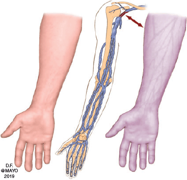
Paget-Schroetter syndrome is rare and it is most often seen in healthy patients in their thirties. It is more common in men and is often reported to occur after vigorous and repetitive activity, due to anatomic abnormalities (e.g. clavicle impingement), or may occur spontaneously.3 Symptoms may include sudden pain, redness, warmth, blue discoloration, and arm swelling4 (Fig. 1).
|
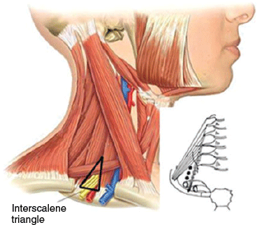
The syndrome is a deep venous thrombosis in an upper extremity, usually in the axillary or subclavian veins, that can lead to a pulmonary embolism. An enlarged hypertrophied anterior scalene muscle can compress the vein posteriorly with further potential for compression by the subclavius muscle. The subclavian vein is extremely vulnerable to injury as it passes through the first rib and clavicle junction, anterior-most to the thoracic outlet (Fig. 2).
|
Cellulitis, lymphangitis and occult malignancies can mimic Paget-Schroetter syndrome and need to be considered in the differential diagnosis. Routine laboratory studies, including a complete blood count, electrolytes, and liver function tests, can be ordered to evaluate for aetiologies of upper extremity oedema. Coagulation studies, including prothrombin time (PT) and partial thromboplastin time (PTT) can be ordered to investigate secondary causes of upper extremity thrombosis.
If Paget-Schroetter syndrome is suspected, the patient should be referred for a duplex ultrasound exam, which is highly accurate in diagnosing this syndrome. The arm is examined in a neutral position since there are thrombi noted at rest in Paget-Schroetter syndrome. Venography is recommended when noninvasive studies are inconclusive, or if intervention is the next step.4 Computed tomography and magnetic resonance imaging have not had the same success in the diagnosis of Paget-Schroetter syndrome due to the absence of structural abnormalities.
Treatment depends on symptom duration and is primarily managed by a vascular surgeon. For symptoms lasting less than 14 days, venography with catheter-directed thrombolysis can be attempted. Catheter-directed thrombolysis is successful in 62% to 84% of cases.5 If thrombolysis is successful, the patient should be referred to a thoracic surgeon for a first rib resection. The patient should receive anticoagulation therapy for 3 to 6 months, followed by repeat sonography. If sonography or venography reveals only a partially-opened vein or residual intrinsic vein defect, a first rib resection should be performed by a thoracic surgeon, with anticoagulation thereafter. A decision can then be made by the vascular surgeon whether to open the vein, stent, repair, and patch, or perform angioplasty.
For symptoms lasting longer than 14 days, venography with catheter-directed thrombolysis should be attempted. If the vein remains completely occluded and there are only mild symptoms, a first rib resection should be performed, along with 3 to 6 months of anticoagulation therapy and repeat sonography. Long-term success rates are 95% to 100%.
Because Paget-Schroetter syndrome is rare and occurs in active individuals prone to other more common conditions, it can often be misdiagnosed. Early recognition and management leads to fewer long-lasting sequelae. Duplex ultrasound is the mainstay for diagnosis. It is imperative to consider this diagnosis in active individuals with upper extremity swelling, and to intervene expeditiously.
Competing interests
The authors declare no conflicts of interest.
Funding source
This research did not receive any specific funding.
Ethical approval
IRB exemption (10/21/2019). IRB exemption was sought and obtained; the article was a case report that used existing documents and records not publicly available. The information obtained did not allow for identification of the patient, either directly or indirectly, and there are no identifiers in the case report. Patient consent was obtained.
References
[1] Sternbach Y, Green RM. Endovascular and surgical management of acute axillary-subclavian venous thrombosis. In: Handbook of venous disease. 2nd Ed. Gloviczki P, Yao JST (Eds). London, UK: Hodder Arnold; (2009) pp. 209–213.[2] Hingorani A, Ascher E, Lorenson E, et al. Upper extremity deep venous thrombosis and its impact on morbidity and mortality rates in a hospital-based population. J Vasc Surg 1997; 26 853–60.
| Upper extremity deep venous thrombosis and its impact on morbidity and mortality rates in a hospital-based population.Crossref | GoogleScholarGoogle Scholar | 9372825PubMed |
[3] Peivandi MT, Nazemian Z. Clavicular fracture and upper-extremity deep venous thrombosis. Orthopedics 2011; 34 227
| Clavicular fracture and upper-extremity deep venous thrombosis.Crossref | GoogleScholarGoogle Scholar | 21410116PubMed |
[4] Di Nisio M, Van Sluis GL, Bossuyt PM, et al. Accuracy of diagnostic tests for clinically suspected upper extremity deep vein thrombosis: a systematic review. J Thromb Haemost 2010; 8 684–92.
| Accuracy of diagnostic tests for clinically suspected upper extremity deep vein thrombosis: a systematic review.Crossref | GoogleScholarGoogle Scholar | 20141579PubMed |
[5] Doyle A, Wolford HY, Davies MG, et al. Management of effort thrombosis of the subclavian vein: today’s treatment. Ann Vasc Surg 2007; 21 723–9.
| Management of effort thrombosis of the subclavian vein: today’s treatment.Crossref | GoogleScholarGoogle Scholar | 17923385PubMed |