

A retrospective review of cutaneous vascular lesions referred to a teledermatology clinic
Amy Choi 1 3 , Amanda Oakley 21 Department of Internal Medicine, Waikato District Health Board, Hamilton, New Zealand; current address: Victoria Clinic, Hamilton, New Zealand
2 Department of Dermatology, Waikato District Health Board, Hamilton, New Zealand
3 Corresponding author. Email: amys.emails.are@gmail.com
Journal of Primary Health Care 13(1) 70-74 https://doi.org/10.1071/HC20046
Published: 18 March 2021
Journal Compilation © Royal New Zealand College of General Practitioners 2021 This is an open access article licensed under a Creative Commons Attribution-NonCommercial-NoDerivatives 4.0 International License
Abstract
INTRODUCTION: Most cutaneous vascular lesions are benign and do not require treatment. Many are referred to specialist dermatologists from primary care.
AIM: This study aimed to investigate the characteristics of cutaneous vascular lesions and the reasons for their referral from primary care.
METHODS: Lesions diagnosed as cutaneous vascular abnormalities or dermatoses were retrospectively selected from a database of patients attending the Waikato Virtual Lesion Clinic. Demographic data, diagnosis and clinic outcome were recorded for each imaged lesion. Primary care referrals were reviewed to determine the reasons for referral.
RESULTS: In total, 229 referrals for vascular lesions were received between January 2010 and February 2019. Patient ages ranged from 6 to 95 years and 64.2% of patients were female. Nearly half the lesions (47.2%) were located on the head and neck; 64.1% had a dermatological diagnosis of a vascular tumour and 18.7% had a malformation. The most common reason for referral was pigmentation (45.7%) and bleeding was least common (8.2%). No diagnosis was given in 34.2% of referrals and less than one-quarter had a correct diagnosis. Malignancy was suspected in 40.2% of referrals; however, the dermatologists found that 95.2% of patients did not require further treatment. Half of excisions (n = 2) were for bleeding and all were histologically benign.
DISCUSSION: Diagnostic uncertainty and suspected malignancy commonly result in referral of benign cutaneous vascular lesions to public dermatology services. This study highlights the usefulness of teledermatology in the timely access of specialist input, minimising the need for intervention or excision.
Keywords: Referrals; cutaneous vascular lesions; primary care; teledermatology
| WHAT GAP THIS FILLS |
| What is already known: We know that cutaneous vascular lesions are common and most of these are benign. Many patients with cutaneous vascular lesions are referred to public teledermatology clinics. |
| What this study adds: This study identifies features of cutaneous vascular lesions that cause diagnostic uncertainly in primary care and demonstrates the usefulness of teledermatology. |
Introduction
Cutaneous vascular lesions are a heterogenous group of disorders involving the blood vessels and lymphatic structures of the skin. They are characterised by dilation, proliferation, aberrant development, or growth of blood vessels.1,2 Although most cutaneous vascular lesions are benign, they may lead to referral to a dermatologist to make or confirm a diagnosis or to confirm that a diagnostic excision is required in primary care.
In 2010, in response to high numbers of referrals to outpatient clinics and an undersupply of dermatologists, the Waikato District Health Board established a teledermatoscopy service known as the Virtual Lesion Clinic in collaboration with MoleMap NZ (Auckland, New Zealand). This was designed to improve access to diagnosis, reduce wait times, and avoid unnecessary interventions.3 Primary care doctors can refer patients with lesions of concern for diagnosis and subsequent treatment (if required). The Virtual Lesion Clinic has a store-and-forward format whereby patients attend a clinic where a melanographer takes a standardised history and regional, macroscopic and dermatoscopic photographs. The files are reviewed by specialist dermatologists who make a diagnosis and provide recommendations that are communicated to patients and their referring clinicians.
The primary objective of this study was to determine the reasons for referral of benign cutaneous vascular lesions from primary care to the Virtual Lesion Clinic. Secondary aims were to classify the lesions and determine the outcomes of the referrals.
Methods
We conducted a retrospective review of cutaneous vascular lesions diagnosed at the Virtual Lesion Clinic between 1 January 2010 and 1 February 2019. All patients had consented in writing to the use of their data and images for research. As the research was observational, Health and Disability Ethics Committee approval was not required.
The lesions were classified as vascular tumours or malformations in accordance with the International Society of the Study of Vascular Anomalies classification system.4 Vascular dermatoses were also included.
Vascular tumours included haemangioma, angiokeratoma, and pyogenic granuloma. Vascular malformations included venous ectasia (venous lake), capillary vascular malformation not otherwise specified (NOS), and vascular naevi. Vascular dermatoses included pigmented purpuric dermatoses (capillaritis and lichen aureus), haemorrhage and ecchymosis. Otherwise unclassified vascular lesions were venous thrombosis and vascular lesion NOS.
Each repeat referral for a vascular lesion within the study timeframe was included as a separate data point. When a patient presented with multiple vascular lesions, each lesion was independently evaluated. Duplicate results were excluded. Demographic details (age, sex and ethnicity), patient characteristics such as Fitzpatrick skin type,5 and the body site of the lesion were recorded.
All referrals were from primary care providers. Common reasons for referral were recorded as: (1) new lesion; (2) growing or changing lesion; (3) raised or nodular lesion; (4) black, pigmented or dark lesion; (5) persistent or non-healing lesion; (6) bleeding lesion. Any provisional diagnosis made by the referrer was also recorded.
The outcome for each cutaneous vascular lesion was: lesion excision, cryotherapy, dermatology clinic assessment, monitor or no action required. When lesion excision was recommended, the corresponding histological report was reviewed.
Results
We found 237 cutaneous vascular lesions in the Virtual Lesion Clinic database within the study timeframe; eight duplicate results were excluded, leaving 229 records for 225 individual lesions in 203 patients.
Patient characteristics
Half of referrals (113, 49.3%) were for patients aged ≥60 years with an age range of 6 – 95 years. There were 147 females (64.2%). European ethnicity was recorded for 214 patients (93%). Māori (10, 4.3%) and Asians (6, 2.6%) were underrepresented in comparison with the 2013 New Zealand Census data6 in which 21.9% of the population of the Waikato region were Māori and 6.9% were Asians.
Lesion characteristics
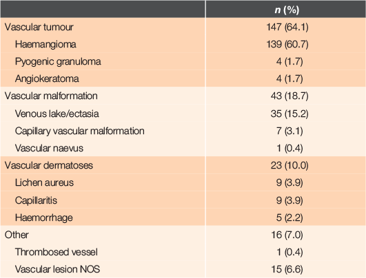
There were 147 vascular tumours (64.1%), primarily haemangioma (139, 60.7%) and 43 vascular malformations (18.7%), mainly venous ectasia (35, 15.2%). Vascular dermatoses comprised 23 referrals (10.0%) and there were 16 unclassified cutaneous vascular lesions (7.0%) (Table 1).
|
One-hundred and eight lesions were located on the head and neck (47.2%), 73 (31.8%) were on the limbs and 48 (21.0%) on the trunk.
Referral characteristics
We reviewed 184 referral letters (80.3% of 229 referrals).
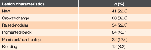
Pigmentation was the most commonly documented characteristic of concern in 84 referrals (45.7%) followed by growth or change in appearance in 60 referrals (32.6%). Bleeding prompted referral in only 15 patients (8.2%) (Table 2). Many referrals included more than one feature of concern.
|
No diagnosis was provided in 63 referrals (34.2% of the 184 referrals reviewed). The most common referral diagnosis was suspected melanoma and non-melanoma skin cancer (74 or 40.2% of referrals). The dermatologist agreed with a referrer’s diagnosis in 36 cases (19.6%). The dermatologist most commonly agreed with the referrer’s diagnosis for haemangiomas (28; this was 20.1% of 139 haemangiomas).
Clinic outcomes
No action was required for 218 lesions (95.2%). Surgical excision was recommended for five lesions (2.2%) and cryotherapy was recommended for one (0.4%). A face-to-face specialist clinic appointment for further assessment was scheduled for five patients (2.2%).
Histology was available for four lesions: two due to recurrent bleeding, one to rule out melanoma and the reason for the other was not stated. All histological reports confirmed the clinical diagnosis of vascular lesions made at the Virtual Lesion Clinic.
Discussion
This study demonstrates the usefulness of teledermatology in the timely diagnosis of cutaneous vascular lesions through improving access to specialist services. Vascular lesions are prevalent in the adult population, yet only ~25 vascular lesions per year were referred to the Virtual Lesion Clinic, indicating that primary care doctors rarely require a specialist opinion. They were usually referred because of diagnostic uncertainty and, in particular, a suspicion of malignancy. Recurrent bleeding and cosmesis were uncommon causes for patient or clinician concern.
A vascular lesion was suspected in only 20% of the referrals. Shared clinical features of cutaneous vascular lesions and malignancies include blue or black discolouration, ulceration, and nodularity.1,7
Training in dermoscopy can improve diagnostic accuracy in primary care and streamline patient care. Dermoscopy has been shown to improve accuracy in the diagnosis of melanoma.7–9 Vascular lesions are straightforward to diagnose with dermoscopy due to their characteristic red or purple patterns (clods (lacunes) or structureless areas) and absence of melanocytic features (eg pigment network).
Angioma appear clinically as a firm papule which is red, purple or blue in colour.1 Under dermoscopy, they are characterised by well-demarcated clods (also called lacunes), dilated vessels and rounded structures. Angiokeratoma is also a red–purple papule with overlying hyperkeratosis. In addition to lacunes it may have a whitish–blue veil, ulceration or rainbow pattern. A pyogenic granuloma is a red papule that grows rapidly and tends to bleed easily, ulcerate and crust. Dermoscopy reveals red homogenous areas, a white collarette, white ‘rail’ lines and ulceration. Venous lake is typically found on the lip and has deep purple, red and blue structureless areas (Table 3).7,10,11

|
Half of the referrals in this study were made for patients aged ≥60 years and most of the referred lesions were located on the head and neck, possibly reflecting the higher incidence of cutaneous malignancies with increasing age and on sun-exposed sites.11,12
Lack of histological confirmation is a limitation of the study, but excisional biopsy for typical benign lesions is unjustified. Avoidance of biopsy improves the patient experience and minimises costs. Specialist advice that a lesion is benign can reassure both referrer and patient, as well as inform future assessment, which is an important advantage of virtual specialist consultations.
Our study has confirmed the value of teledermatoscopy for vascular lesions.
Competing interests
Amanda Oakley is a consultant for MoleMap NZ and is paid a fee for service in the private sector. Amy Choi has no conflicts of interest to declare.
Acknowledgements
We thank MoleMap New Zealand for providing the database and images. This research did not receive any specific funding.
References
[1] Wirth FA, Lowitt MH. Diagnosis and treatment of cutaneous vascular lesions. Am Fam Physician. 1998; 57 765–73.| 9490999PubMed |
[2] Patel AM, Chou EL, Findeiss L, Kelly K. The horizon for treating cutaneous vascular lesions. Semin Cutan Med Surg. 2012; 31 98–104.
| The horizon for treating cutaneous vascular lesions.Crossref | GoogleScholarGoogle Scholar | 22640429PubMed |
[3] Lim D, Rademaker M, Oakley A. Better, sooner, more convenient – a successful teledermoscopy service. Australas J Dermatol. 2012; 53 22–5.
| Better, sooner, more convenient – a successful teledermoscopy service.Crossref | GoogleScholarGoogle Scholar | 22309326PubMed |
[4] Wassef M, Blei F, Adams D, et al. Vascular anomalies classification: recommendations from the International Society for the Study of Vascular Anomalies. Pediatrics. 2015; 136 e203–14.
| Vascular anomalies classification: recommendations from the International Society for the Study of Vascular Anomalies.Crossref | GoogleScholarGoogle Scholar | 26055853PubMed |
[5] Fitzpatrick TB. The validity and practicality of sun-reactive skin types i through vi. Arch Dermatol. 1988; 124 869–71.
| The validity and practicality of sun-reactive skin types i through vi.Crossref | GoogleScholarGoogle Scholar | 3377516PubMed |
[6] Statistics New Zealand. 2013 Census quickstats about a place: Waikato region [Internet]. Wellington: Stats NZ; 2013. [cited 2019 December 3]. Available from: http://archive.stats.govt.nz
[7] Piccolo V, Russo T, Moscarella E, et al. Dermatoscopy of vascular lesions. Dermatol Clin. 2018; 36 389–95.
| Dermatoscopy of vascular lesions.Crossref | GoogleScholarGoogle Scholar | 30201148PubMed |
[8] Congalton AT, Oakley AM, Rademaker M, et al. Successful melanoma triage by a virtual lesion clinic (teledermatoscopy). J Eur Acad Dermatol Venereol. 2015; 29 2423–8.
| Successful melanoma triage by a virtual lesion clinic (teledermatoscopy).Crossref | GoogleScholarGoogle Scholar | 26370585PubMed |
[9] Vestergaard ME, Macaskill P, Holt PE, Menzies SW. Dermoscopy compared with naked eye examination for the diagnosis of primary melanoma: a meta-analysis of studies performed in a clinical setting. Br J Dermatol. 2008; 159 669–76.
| Dermoscopy compared with naked eye examination for the diagnosis of primary melanoma: a meta-analysis of studies performed in a clinical setting.Crossref | GoogleScholarGoogle Scholar | 18616769PubMed |
[10] Lee JS, Mun JH. Dermoscopy of venous lake on the lips: a comparative study with labial melanotic macule. PLoS One. 2018; 13 e0206768
| Dermoscopy of venous lake on the lips: a comparative study with labial melanotic macule.Crossref | GoogleScholarGoogle Scholar | 30592769PubMed |
[11] Ouyang YH. Skin cancer of the head and neck. Semin Plast Surg. 2010; 24 117–26.
| Skin cancer of the head and neck.Crossref | GoogleScholarGoogle Scholar | 22550432PubMed |
[12] Environmental Health Indicators. Melanoma deaths. [Factsheet]. Wellington: Environmental Health Indicators Programme, Massey University; 2019.