

Q fever: pets, vets and validating tests
Jacqueline M Norris A C , Katrina L Bosward A and Jane Heller BA Faculty of Veterinary Science
The University of Sydney
NSW 2006, Australia
B School of Animal and Veterinary Sciences
Charles Sturt University
Locked Bag 588
Wagga Wagga, NSW 2678, Australia
C Tel: +61 2 9351 7095, Email: jacqui.norris@sydney.edu.au
Microbiology Australia 34(4) 186-188 https://doi.org/10.1071/MA13064
Published: 18 September 2013
Q fever is a highly significant worldwide zoonosis caused by the bacterium Coxiella burnetii. While infection is commonly asymptomatic, 40% of primary infections in humans are symptomatic, with serious acute or chronic debilitating illnesses possible, including endocarditis, post-Q fever fatigue syndrome and recrudescent granulomatous lesions in bone or soft tissue1,2. The bacterium itself has a duplicitous lifecycle; a metabolically active form obligately replicates within the macrophage cell lineage while an inactive form has extreme environmental resilience, providing a means to travel to new cells and new hosts. Coupled with a potentially large, seemingly asymptomatic reservoir encompassing wild and domestic mammals, birds and arthropods, this bacterium continues to raise important questions about its impact on public health worldwide. Given the targeted and complex nature of testing required to confirm a diagnosis in humans3, ongoing vigilance in promptly recognising clinical cases in humans and reappraisal of the potential risks created by animal exposure is required. This article outlines the current evidence on the potential role that cats and dogs might play in transmission of this bacterium and provides a framework for future studies.
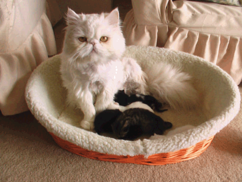
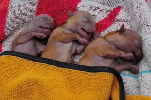
Q fever has been traditionally framed as an occupational disease, associated with contact with cattle, sheep and goats in the livestock and meat industries. This has been both a help and a hindrance in identifying human cases and accurately assessing risk of exposure to C. burnetii. However, isolated reports of community-acquired Q fever have highlighted the potential role of pet dogs and cats as sources of this pathogen4–11. Most reported cases have been associated with direct or indirect exposure to breeding queens or bitches and/or neonatal kittens (Figure 1) or puppies (Figure 2) during or shortly after parturition. Given the large number of bacteria in placental tissue reported in other animal species1,12, contact with birth product contaminated animals, surfaces or aerosols is the most likely source of the pathogen in these cases. The reports of feline or canine associated Q fever cases in humans are infrequent given the millions of cats and dogs worldwide, which might imply low carriage rates in these animal species. Supporting this is the finding that cat and dog ownership is not associated with increased seroprevalence of antibodies to C. burnetii13. However, the consequence of Q fever for those individuals at risk, such as unvaccinated veterinary personnel or breeders of pets, might be severe8,10. Therefore, a broad and coordinated approach is required for future research from medical and veterinary clinicians and researchers in order to raise the profile of Q fever as a diagnostic consideration.
|
|
Determining C. burnetii infection in dogs and cats has been complicated by the lack of confirmed disease associations and the absence of standardised sensitive and specific diagnostic techniques. Serological tests, on which diagnosis primarily relies, cannot be translated directly from one species to another without standardisation14. The World Organisation for Animal Health (OIE) reference test for serological diagnosis of C. burnetii remains the complement fixation tests (CFT)15. Although CFT is highly specific, indirect immunofluorescence assay (IFA) and enzyme-linked immunosorbent assay (ELISA) are frequently used in human medicine due to their higher sensitivity, earlier detection of seroconversion and comparative ease to perform1,14. The standardisation of serological tests for determining C. burnetii exposure in cats and dogs has been hampered to some extent by the absence of clearly identified clinical disease associations in these animal species (limiting the ability to define true positives and negatives) and the dangers of laboratory culture of this bacterium, which requires PC3 facilities. The tendency of many researchers looking at the broader question of the prevalence of infection in dog and cat populations therefore has been to extrapolate diagnostic testing methods and cut-off points used for human serum or to use positive and negative controls from non-canine or non-feline species, which raises questions over the reliability of results.
The reports of isolated outbreaks of community-acquired Q fever related to dog or cat contact has stimulated opportunistic seroprevalence studies searching for answers as to how widespread infection of cats and dogs might be. In maritime Canada, exposure to parturient cats and newborn kittens has been identified as a significant risk factor for Q fever16, with seroprevalence of C. burnetii infection in cats in these regions varying from 6.2 to 32%17–19. In other countries (such as South Africa, Japan and the USA), seroprevalence has ranged from 1.9 to 42%20–23. However, the serological testing methods varied between studies from IFA using phase II or both phase I and II C. burnetii antigen (Nine Mile strain) to micro agglutination assays with cut-off values for either test ranging from 1/4 to 1/64 serum dilutions with explanation of positive and negative controls insufficient to determine the choice of cut-off value. In contrast, some studies have cross checked results from methods such as IFA with the OIE standard of CFT and used serum samples from cats at the centre of a Q fever outbreak in humans to determine positive controls8,19. Thus, comparison of results of differing studies should not be performed without knowledge and critique of the methods used.
Seroprevalence studies in dogs have produced variable results (0–35%) using ELISA24 or IFA25–29 from dogs sampled in Canada, Italy, Egypt, France and French colonies, Australia and post-Iraq military deployment dogs from the USA. Again, the variation in methods used, explanation (or absence) of positive and negative controls, description of sample population and determination of cut-off values makes comparison between studies difficult and determination of real prevalence in dog populations complex from these data.
Recently, molecular methods used to determine the presence of C. burnetii DNA on healthy vaginal or uterine tissues of healthy cats pre- or postdesexing in Colorado, USA, found 4/47 (8.5%) pet cats had evidence of C. burnetii DNA30. While in the Netherlands following the 2007–2010 Q fever outbreak, C. burnetii DNA was not detected in placentas from cats (n = 15), but was found in 4/54 (7%) dog placentas derived from veterinary clinical practices focussed on breeding pets31.
Future research into the potential role that our closest companions might play in Q fever needs to take a broader population perspective, comparing the incidence of Q fever and prevalence of prior infection in subpopulations of potentially at-risk people, such as veterinary personnel and dog and cat breeders, with the broader Australian population. From a canine and feline perspective, further refinement and standardisation of serological assays and molecular methods against OIE standards is required to determine the prevalence of asymptomatic infection, persistence of infection and to explore potentially unrecognised disease associations and risk factors within subpopulations of the stray, feral, pet and breeding cat and dog subpopulations.
References
[1] Maurin, M. and Raoult, D. (1999) Q fever. Clin. Microbiol. Rev. 12, 518–553.| 1:STN:280:DyaK1MvksFCnsw%3D%3D&md5=55cf2cf5c2628ca47705399e30b29358CAS | 10515901PubMed |
[2] Raoult, D. et al. (2005) Natural history and pathophysiology of Q fever. Lancet Infect. Dis. 5, 219–226.
| Natural history and pathophysiology of Q fever.Crossref | GoogleScholarGoogle Scholar | 1:STN:280:DC%2BD2M7mtFShsw%3D%3D&md5=5434b4feff88e351eae0ff5335d8e22bCAS | 15792739PubMed |
[3] Porter, S.R. et al. (2011) Q fever: current state of knowledge and perspectives of research of a neglected zoonosis. Int. J. Microbiol. 2011, 248418.
| Q fever: current state of knowledge and perspectives of research of a neglected zoonosis.Crossref | GoogleScholarGoogle Scholar | 22194752PubMed |
[4] Buhariwalla, F. et al. (1996) A dog-related outbreak of Q fever. Clin. Infect. Dis. 23, 753–755.
| A dog-related outbreak of Q fever.Crossref | GoogleScholarGoogle Scholar | 1:STN:280:DyaK2s%2Fms1emug%3D%3D&md5=33036884ae9cc664c4a22134993f2184CAS | 8909839PubMed |
[5] Kosatsky, T. (1984) Household outbreak of Q-fever pneumonia related to a parturient cat. Lancet 324, 1447–1449.
| Household outbreak of Q-fever pneumonia related to a parturient cat.Crossref | GoogleScholarGoogle Scholar |
[6] Marrie, T.J. et al. (1989) Truckin’ pneumonia: an outbreak of Q fever in a truck repair plant probably due to aerosols from clothing contaminated by contact with newborn kittens. Epidemiol. Infect. 102, 119–127.
| Truckin’ pneumonia: an outbreak of Q fever in a truck repair plant probably due to aerosols from clothing contaminated by contact with newborn kittens.Crossref | GoogleScholarGoogle Scholar | 1:STN:280:DyaL1M7jslartw%3D%3D&md5=dcc5e74e39643f8db182019f5def1e48CAS | 2917613PubMed |
[7] Marrie, T.J. et al. (1988) An outbreak of Q fever probably due to contact with a parturient cat. Chest 93, 98–103.
| An outbreak of Q fever probably due to contact with a parturient cat.Crossref | GoogleScholarGoogle Scholar | 1:STN:280:DyaL1c%2FotFyisg%3D%3D&md5=0ead9b44f0bb5ff2a9d66ca0c5f7c328CAS | 3335174PubMed |
[8] Kopecny, L. et al. (2013) Investigating Coxiella burnetii infection in a breeding cattery at the centre of a Q fever outbreak. J. Feline Med. Surg. , 7 May 2013.
[9] Langley, J.M. et al. (1988) Poker players pneumonia – an urban outbreak of Q fever following exposure to a parturient cat. N. Engl. J. Med. 319, 354–356.
| Poker players pneumonia – an urban outbreak of Q fever following exposure to a parturient cat.Crossref | GoogleScholarGoogle Scholar | 1:STN:280:DyaL1c3ns1Srtg%3D%3D&md5=2b607c7d119c74ec9b6736eb31b47076CAS | 3393197PubMed |
[10] Maywood, P. (2011) Outbreak investigation: Q Fever in a small animal hospital. Australian College of Veterinary Scientists.
[11] Pinsky, R.L. et al. (1991) An outbreak of cat-associated Q fever in the United States. J. Infect. Dis. 164, 202–204.
| An outbreak of cat-associated Q fever in the United States.Crossref | GoogleScholarGoogle Scholar | 1:STN:280:DyaK3M3nslCktQ%3D%3D&md5=9b381b81f5d7b312b8a97ff2bd2fcbedCAS | 2056206PubMed |
[12] Babudieri, B. (1959) Q fever: a zoonosis. Adv. Vet. Sci. 5, 81–182.
[13] Skerget, M. et al. (2003) Cat or dog ownership and seroprevalence of ehrlichiosis, Q fever, and cat-scratch disease. Emerg. Infect. Dis. 9, 1337–1340.
| Cat or dog ownership and seroprevalence of ehrlichiosis, Q fever, and cat-scratch disease.Crossref | GoogleScholarGoogle Scholar | 14609477PubMed |
[14] Fournier, P.E. et al. (1998) Diagnosis of Q fever. J. Clin. Microbiol. 36, 1823–1834.
| 1:STN:280:DyaK1czht12nsg%3D%3D&md5=bc9916cc05949111fa68e27d9b0766f6CAS | 9650920PubMed |
[15] Rousset, E. et al. (2010) Q Fever. In Manual of Diagnostic Tests and Vaccines for Terrestrial Animals, pp. 292–303, Paris, Office International des Epizooties (OIE).
[16] Marrie, T.J. et al. (1988) Exposure to parturient cats - a risk factor for acquisition of Q fever in maritime Canada. J. Infect. Dis. 158, 101–108.
| Exposure to parturient cats - a risk factor for acquisition of Q fever in maritime Canada.Crossref | GoogleScholarGoogle Scholar | 1:STN:280:DyaL1c3nsVSktg%3D%3D&md5=4d67162831b9760cac7a716ff4e85445CAS | 3392409PubMed |
[17] Higgins, D. and Marrie, T.J. (1990) Seroepidemiology of Q fever among cats in New Brunswick and Prince Edward Island. Ann. N. Y. Acad. Sci. 590, 271–274.
| Seroepidemiology of Q fever among cats in New Brunswick and Prince Edward Island.Crossref | GoogleScholarGoogle Scholar | 1:STN:280:DyaK3czjsVegsw%3D%3D&md5=6d1d6685b59fdf6bab8e6b8005e60727CAS | 2378456PubMed |
[18] Marrie, T.J. et al. (1985) Seroepidemiology of Q fever among domestic animals in Nova Scotia. Am. J. Public Health 75, 763–766.
| Seroepidemiology of Q fever among domestic animals in Nova Scotia.Crossref | GoogleScholarGoogle Scholar | 1:STN:280:DyaL2M3it1ygsg%3D%3D&md5=76e3ab2929531d5c65312df919075d00CAS | 3890569PubMed |
[19] Vallieres, A. et al. (1996) Seroprevalence of Coxiella burnetii within a domestic cat population in Quebec. In Animal Epidemiology and Protection of Public Health, pp. 43–49.
[20] Matthewman, L. et al. (1997) Exposure of cats in southern Africa to Coxiella burnetii, the agent of Q fever. Eur. J. Epidemiol. 13, 477–479.
| Exposure of cats in southern Africa to Coxiella burnetii, the agent of Q fever.Crossref | GoogleScholarGoogle Scholar | 1:STN:280:DyaK2svgtlWrtQ%3D%3D&md5=03240d745891060a238ee42a8eece42aCAS | 9258556PubMed |
[21] Komiya, T. et al. (2003) Seroprevalence of Coxiella burnetii infections among cats in different living environments. J. Vet. Med. Sci. 65, 1047–1048.
| Seroprevalence of Coxiella burnetii infections among cats in different living environments.Crossref | GoogleScholarGoogle Scholar | 14532705PubMed |
[22] Morita, C. et al. (1994) Seroepidemiological survey of Coxiella burnetii in domestic cats in Japan. Microbiol. Immunol. 38, 1001–1003.
| 1:CAS:528:DyaK2MXjtFaqtrs%3D&md5=009ec5c86df7fe31637fdc3991ff2f53CAS | 7723682PubMed |
[23] Willeberg, P. et al. (1980) Environmental exposure to Coxiella burnetii – a sero-epidemiologic survey among domestic animals. Am. J. Epidemiol. 111, 437–443.
| 1:STN:280:DyaL3c7psFKlug%3D%3D&md5=50b23f5fe8f3caa963f6a2f4442c3915CAS | 7377186PubMed |
[24] Cooper, A. et al. (2011) Serological evidence of Coxiella burnetii infection in dogs in a regional centre. Aust. Vet. J. 89, 385–387.
| Serological evidence of Coxiella burnetii infection in dogs in a regional centre.Crossref | GoogleScholarGoogle Scholar | 1:STN:280:DC%2BC3MfltFGntA%3D%3D&md5=62b930bc07ef58d84b5c05fc3da0a135CAS | 21933165PubMed |
[25] Marrie, T.J. et al. (1985) Seroepidemiology of Q fever among domestic animals in Nova Scotia. Am. J. Public Health 75, 763–766.
| Seroepidemiology of Q fever among domestic animals in Nova Scotia.Crossref | GoogleScholarGoogle Scholar | 1:STN:280:DyaL2M3it1ygsg%3D%3D&md5=76e3ab2929531d5c65312df919075d00CAS | 3890569PubMed |
[26] Amal, S.M.S. et al. (2002) Prevalence of Coxiella burnetii infection among dogs and humans in upper Egypt. Assiut Veterinary Medical Journal 47, 205–215.
[27] Boni, M. et al. (1998) Survey of seroprevalence of Q fever in dogs in the southeast of France, French Guyana, Martinique, Senegal and the Ivory Coast. Vet. Microbiol. 64, 1–5.
| Survey of seroprevalence of Q fever in dogs in the southeast of France, French Guyana, Martinique, Senegal and the Ivory Coast.Crossref | GoogleScholarGoogle Scholar | 1:STN:280:DyaK1M%2FovFCqtg%3D%3D&md5=f7b3f2e1e70c8fc70bf9a3de6124494eCAS | 9874098PubMed |
[28] Havas, K.A. and Burkman, K. (2011) A comparison of the serological evidence of Coxiella burnetii exposure between military working dogs and feral canines in Iraq. Mil. Med. 176, 1101–1103.
| 22128642PubMed |
[29] Baldelli, R. et al. (1992) Dog-transmitted zoonoses: a serological survey in the province of Bologna. Ann. Ist. Super. Sanita 28, 493–496.
| 1:STN:280:DyaK3s3mtlCiug%3D%3D&md5=1d34abedf312939f3c0614af737a3731CAS | 1303042PubMed |
[30] Cairns, K. et al. (2007) Prevalence of Coxiella burnetii DNA in vaginal and uterine samples from healthy cats of north-central Colorado. J. Feline Med. Surg. 9, 196–201.
| Prevalence of Coxiella burnetii DNA in vaginal and uterine samples from healthy cats of north-central Colorado.Crossref | GoogleScholarGoogle Scholar | 17208030PubMed |
[31] Roest, H.I.J. et al. (2013) Search for possible additional reservoirs for human Q fever, The Netherlands. Emerg. Infect. Dis. 19, 834–835.
| Search for possible additional reservoirs for human Q fever, The Netherlands.Crossref | GoogleScholarGoogle Scholar |
Biographies
Dr Jacqueline Norris is an Associate Professor in Veterinary Microbiology at the Faculty of Veterinary Science, The University of Sydney. Her research interests include: new diagnostic and therapeutic approaches to feline viral diseases such as those caused by feline infectious peritonitis virus, feline leukemia virus and feline immunodeficiency virus; aetiology and diagnostic testing for renal disease in domestic and non-domestic felids; antimicrobial resistance in zoonotic pathogens; Q fever and the epidemiology, diagnosis and disease outcomes of Coxiella infections in animals.
Dr Katrina Bosward is a Senior Lecturer in Veterinary Microbiology at the Faculty of Veterinary Science, The University of Sydney. Her research interests include Q fever and Coxiella burnetii – its diagnosis and prevention both in humans and animals; infectious diseases of dairy cattle especially Streptococcal and Staphylococcal mastitis and those caused by Mycoplasma spp.
Dr Jane Heller is a Senior Lecturer in Veterinary Epidemiology and Public Health in the School of Animal and Veterinary Science, Charles Sturt University. Her research interests focus on infectious disease epidemiology, with particular reference to the potential for zoonotic transfer of pathogens between animals and humans. Her current areas of research include: E. coli O157 shedding in cattle; risk factors for Q fever exposure; defining the extent of understanding of evidence-based medicine in students and teachers and identification of neoplastic causes of cattle carcase condemnation. She is an associate member of the Epidemiology Chapter of the Australian College of Veterinary Scientists and a Diplomate of the European College of Veterinary Public Health, in the subspecialty of population medicine.