Advances in the understanding of the Cryptococcus neoformans and C. gattii species complexes and cryptococcosis
Carolina Firacative A C , Luciana Trilles B and Wieland Meyer A BA Molecular Mycology Research Laboratory, CIDM, Sydney Medical School – Westmead Hospital, MBI, The University of Sydney, WIMR, Sydney, Australia
B Laboratório de Micologia, Instituto Nacional de Infectologia Evandro Chagas (INI), Fundação Oswaldo Cruz (FIOCRUZ), Rio de Janeiro, Brazil
C Tel: +61 2 8627 3432, Email: sfir4568@uni.sydney.edu.au
Microbiology Australia 38(3) 106-111 https://doi.org/10.1071/MA17043
Published: 11 August 2017
The rising incidence of cryptococcosis, a potentially fatal fungal infection affecting both immunocompromised and immunocompetent humans and animals, and the emergence of disease outbreaks, has increased the need for more in-depth studies and constant vigilance of its two etiological agents, the cosmopolitan and well known Cryptococcus neoformans and its sibling species C. gattii. As a result, a global scientific network has established formal links between institutions to gain better insights into Cryptococcus and cryptococcosis, enabling collaborations amongst researchers with different backgrounds, perspectives and skills. Interdisciplinary projects include: (1) the study of the ecology and geographical distribution of the agents of cryptococcosis; (2) the application of new alternative methodologies for the rapid and accurate identification of the two sibling species and major molecular types/possible cryptic species (VNI-VNIV and VGI-VGIV); (3) the use of different animal models of infection to assess cryptococcal pathogenesis and virulence factors; and (4) population genetics studies directed towards the discovery of virulence/tissue tropism associated genetic signatures. These studies enrich the knowledge and understanding of the epidemiology of this mycosis and help to better comprehend fungal virulence, genetics, pathogenesis, antifungal susceptibility, as well as investigating the regional and global spread, to improve treatment options of the disease caused by these important emerging pathogenic yeasts.
The encapsulated basidomycetes yeasts C. neoformans and C. gattii are the causative agents of cryptococcosis worldwide, causing pneumonia and meningoencephalitis in both, immunocompetent and immunocompromised hosts, resulting in a high morbidity and mortality. With almost 250 000 people affected with cryptococcal meningitis per year, this fungal infection is still responsible for more than 180 000 deaths anually1. By combining data from different studies carried out in numerous laboratories from around the world, it has been possible to define the geographical distribution of human clinical, animal and environmental cryptococcal isolates and to characterise different aspects of their biology.
Taxonomy, epidemiology and ecology
Within the currently accepted two species, four serotypes, distinguished by the capsular polysaccharide, and eight major molecular types, which are possible cryptic species, have been recognised. These major molecular types have been identified by different genotyping methodologies including PCR fingerprinting2, amplified fragment length polymorphisms (AFLP)3, restriction fragment length polymorphisms (RFLP)4 and multilocus sequence typing (MLST)5. In C. neoformans the serotype A, comprises the molecular types VNI (VNB) and VNII, the serotype D, molecular type VNIV, and the AD hybrid between serotypes A and D, molecular type VNIII. In C. gattii the serotypes B and C comprise both the molecular types VGI, VGII, VGIII and VGIV. From these, seven molecular types have recently been proposed to be raised to species level6, although the issue of species definition is still controversial amongst the cryptococcal research community, resulting in the recognition of C. neoformans and C. gattii as species complexes7.
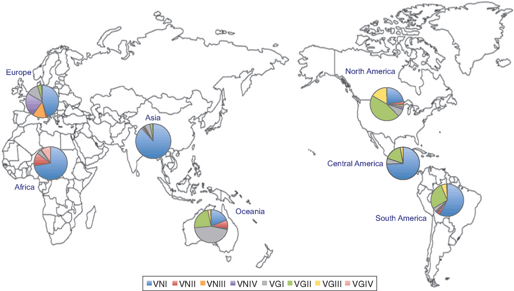
Epidemiological studies have shown that C. neoformans molecular type VNI causes the majority of the cases of cryptococcosis and that this species is also most commonly isolated from the environment worldwide, except in Australia and Papua New Guinea, where VGI is the most common type8–10. VNII is an uncommon molecular type, which is reported from all the continents but in very low percentages9,11. VNIII AD hybrids are also recognised but their frequency seems to be strictly related to the presence of VNIV molecular type. In Europe and in the USA, where the frequency of isolation of VNIV strains is higher than in other geographical areas, a similar percentage of VNIII isolates has been observed, suggesting that in these regions hybridisation between the haploid VNI and VNIV populations is occurring9,11. Although C. gattii is less frequently recovered and it was considered to be geographically restricted to tropical and sub-tropical regions of the world, new studies have reported its extent to temperate regions and additional ecological reservoirs12–16. Besides the molecular type VGII isolates reported extensively from the Vancouver Island outbreak and its expansion to the Pacific Northwest of the USA16,17, this molecular type has also been reported from Brazil, Colombia and Australia, with some cases in Europe10–12,15. The molecular type VGIII has mostly been recovered from Mexico, Colombia and the USA11,13,18, while it is very rare in other continents11. Apart from Oceania, VGI is found in Asia, North, Central and South America, and Europe, where it counts for less than 10% of the isolates and is rare in Africa9. The molecular type VGIV has been reported from Southern Africa, India, Colombia and Mexico9,11 (Figure 1).

|
Unlike C. neoformans, which predominantly affects immunocompromised hosts, C. gattii most often affects patients with no apparent risk factors1,18–21. In addition, in both species, the same genotypes have been found to be recovered from patients and from the patients’ environment19,22, which supports the theory of the acquisition of the infection from environmental sources and reaffirms the saprophytic and arboreal sources of these yeasts. Both species have also been isolated frequently from air, and swabs from trees, from decaying wood in hollows of many tree species, either individually or together14,23. Regarding the epidemiology of clinical cases of cryptococcosis, male patients are more commonly reported, as it is thought that male immune response may be less efficient in controlling cryptococcal infection24, whilst cases in children and older patients are still rare in most countries18,19. In domestic animals from North America, C. neoformans molecular type VNI has been found to affect mostly dogs whereas C. gattii molecular type VGIII predominantly infects cats. Genotypic analysis of such veterinary cases found as well that they are closely related to human and environmental cases and that VGII isolates presented higher minimum inhibitory concentrations (MICs) for most antifungal drugs than C. neoformans and other molecular types in C. gattii25.
Molecular type and species identification
Between and within the major molecular types/species, there are differences in the ecology, epidemiology, clinical manifestations and antifungal drugs susceptibility that emphasise the need for rapid and accurate differentiation of Cryptococcus spp. Two methodologies have been assessed and adopted as a possible alternative for a distinction of the species and major molecular types of C. neoformans and C. gattii, compared with the currently used conventional techniques, such as PCR fingerprinting, AFLP and RFLP. Matrix-assisted laser desorption ionisation time of flight mass spectrometry (MALDI-TOF MS) has been shown to be a suitable tool for inter- and intra-specific differentiation of human, animal and environmental strains of both cryptococcal species and also allowed for the detection of hybrid strains (Figure 2)26. As a result of this study, an in-house spectral library was constructed with the obtained spectra made available for use in a clinical diagnostic laboratory setting. Given the high costs of the initial setup of MALDI-TOF MS, a hyper-branched rolling circle PCR (HRCA) assay was developed as a substitute, a fast and specific method applicable in resource restricted countries. This method, which is based on the detection of specific single nucleotide polymorphisms in the phospholipase B gene among all major molecular types, proved to be fast, specific and highly sensitive and reproducible, for the differentiation of the molecular types and identification of hybrids within C. neoformans and C. gattii from human clinical samples (Figure 2)27.
Animal models for the study of cryptococcosis
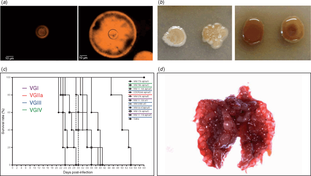
High (VGIIa and VGIIc) and low (VGIIb) virulent subtypes within C. gattii molecular type VGII are associated with outbreaks of infection in British Columbia, Canada and the Pacific Northwest of the USA16,17 but less is known about the virulence of the major molecular types of C. gattii. A number of animal models, including a mouse and two invertebrate models of infection, have been used to determine whether or not there is a relationship between the molecular type and the virulence of the strains. The mouse model is a very well established system to study virulence and uses inoculation via the respiratory route. However, Drosophila melanogaster and larvae of the wax moth Galleria mellonella are gaining prominence as alternative models to study pathogenesis, given that insects can be used and maintained in large quantities with low costs, are easy to handle and their utilisation does not require ethical permission. In addition, fungal virulence factors involved in mammalian pathogenesis, such as capsule enlargement and melanin production, are also important during infection and death of insects (Figure 3a, b). In a D. melanogaster model, VGIII was found to be the most virulent molecular type when grown at 30°C, while VGII had the higher MICs against fluconazole and VNI grew most rapidly at 37°C, synthesised more melanin at 30°C and was more resistant to H2O2. This model showed, however, that temperature played a significant role in the flies’ survival28. In a G. mellonella model, performed at 37°C, no differences amongst the major molecular types were observed in larvae survival and the expression of virulence factors, including capsule production, melanin synthesis and growth rate at 37°C. However, isolates with enhanced virulence were recognised among VGI, VGIII and VGIV, which importantly suggests that virulence might be associated with specific attributes of the strains that need further characterisation29, and which may not be associated with the temperature at which the experiments are carried out in the different model systems. After intranasal infection of Balb/C mice with the highly virulent strains identified in the insect models, comparable results were found and, in addition, formation of multiple granulomas in the lungs was observed, which supported pulmonary cryptococcosis as the main outcome of infection (Figure 3c, d).
Whole genome sequencing-based population genetics to discover genetic differences relevant to pathogenicity and tissue tropism
In order to detect speciation and genetic differences within the distinct emergent populations of C. gattii, whole genome sequencing (WGS) has been performed on 134 C. gattii VGII isolates from five continents, including the subtypes VGIIa, VGIIb and VGIIc16,17, which were previously studied by MLST analysis. Although the VGII subtypes were found to be completely clonal, the overall VGII population showed a high genetic diversity. In addition, several mutations, deletions, transpositions and recombination events have been identified within VGII strains, and this pointed to a South American origin30. Using WGS to compare subtypes, various genetic differences have been recognised, which are potentially related to habitat adaptation, virulence, and pathology30. For example, a set of ~50 genes has been identified being either present or absent in lung or brain infecting strains30, offering the possibility to identify biomarkers, which should be able to guide clinical treatment to reduce mortality in patients based on the early detection of strains with a specific tissue tropism.
Similarly, to gain insights in the emerging C. gattii molecular type VGIII, more than a hundred isolates from endemic areas and sporadic cases have been characterised. The VGIII population was found to have a high genetic diversity but clustered into two main groups, which correspond with either serotype B or C. WGS revealed Mexico and the USA as a likely origin of the serotype B VGIII population, and Colombia as a possible origin of the serotype C VGIII population. Importantly, serotype B isolates were found to be more virulent than serotype C isolates in a mouse model of infection, while serotype C showed to be less susceptible to fluconazole and azoles than serotype B isolates. These results emphasise the importance of the molecular characterisation of the isolates causing cryptococcosis in order to choose an appropriate and timely treatment for cryptococcal infection22.
In conclusion, the rising occurrence of cryptococcosis in humans and animals, the emergence of outbreaks and the decreased susceptibility to the commonly used antifungal drugs, and the expansion of the ecological niche of cryptococcal species highlight the importance of a constant interaction between researchers with different backgrounds, perspectives and skills, enabling increased vigilance of these pathogenic yeasts. Differences regarding the epidemiology, virulence, and antifungal susceptibility between and within species are relevant to disease outcome and fungal therapy. Thus, the correct and fast recognition of the major molecular types within C. neoformans and C. gattii and their phenotypic and genotypic characterisation is essential, as it will most certainly decrease the time from diagnosis to treatment, to lead to a more effective treatment and reduced mortality rates. Continuous collaborative studies on the ecology, genetics and pathogenesis of these medically important yeasts are necessary to contribute, integrate and enrich our knowledge of this important emerging mycosis.
Acknowledgements
This work results as part of the continuous efforts and collaboration among multiple institutions and research groups from around the world, working together and devoted to study Cryptococcus and cryptococcosis. It was supported by grants from the National Health and Medical Research Council (NHMRC) of Australia (APP1031943), The National Institute of Health (NIH) USA (R21AI098059), and the program science without borders (CAPES) Brazil (098/2012) to WM and by the Coordenação de Aperfeiçoamento de Pessoal de Nível Superior’ (CAPES) of Brazil (098/2012) to LT.
References
[1] Rajasingham, R. et al. (2017) Global burden of disease of HIV-associated cryptococcal meningitis: an updated analysis. Lancet Infect. Dis. 17, 873–888.| Global burden of disease of HIV-associated cryptococcal meningitis: an updated analysis.Crossref | GoogleScholarGoogle Scholar |
[2] Meyer, W. and Mitchell, T.G. (1995) Polymerase chain reaction fingerprinting in fungi using single primers specific to minisatellites and simple repetitive DNA sequences: strain variation in Cryptococcus neoformans. Electrophoresis 16, 1648–1656.
| Polymerase chain reaction fingerprinting in fungi using single primers specific to minisatellites and simple repetitive DNA sequences: strain variation in Cryptococcus neoformans.Crossref | GoogleScholarGoogle Scholar | 1:CAS:528:DyaK2MXovV2ltrw%3D&md5=262fbea364005c075063e4b6d9f594d8CAS |
[3] Boekhout, T. et al. (2001) Hybrid genotypes in the pathogenic yeast Cryptococcus neoformans. Microbiology 147, 891–907.
| Hybrid genotypes in the pathogenic yeast Cryptococcus neoformans.Crossref | GoogleScholarGoogle Scholar | 1:CAS:528:DC%2BD3MXjtVeisLw%3D&md5=a65234f2f91f1fbf14957dceb7806f7eCAS |
[4] Meyer, W. et al. (2003) Molecular typing of IberoAmerican Cryptococcus neoformans isolates. Emerg. Infect. Dis. 9, 189–195.
| Molecular typing of IberoAmerican Cryptococcus neoformans isolates.Crossref | GoogleScholarGoogle Scholar |
[5] Meyer, W. et al. (2009) Consensus multi-locus sequence typing scheme for Cryptococcus neoformans and Cryptococcus gattii. Med. Mycol. 47, 561–570.
| Consensus multi-locus sequence typing scheme for Cryptococcus neoformans and Cryptococcus gattii.Crossref | GoogleScholarGoogle Scholar | 1:CAS:528:DC%2BD1MXht1agu7bF&md5=c71dd4b04f29fc35fded676a93f46a6eCAS |
[6] Hagen, F. et al. (2015) Recognition of seven species in the Cryptococcus gattii/Cryptococcus neoformans species complex. Fungal Genet. Biol. 78, 16–48.
| Recognition of seven species in the Cryptococcus gattii/Cryptococcus neoformans species complex.Crossref | GoogleScholarGoogle Scholar | 1:CAS:528:DC%2BC2MXjs1ajtr8%3D&md5=f195b8021d2cf9dc0da07d2cb3979c71CAS |
[7] Kwon-Chung, K.J. et al. (2017) The case for adopting the ‘species complex’ nomenclature for the etiological agents of cryptococcosis. MSphere 2, e00357-16.
| The case for adopting the ‘species complex’ nomenclature for the etiological agents of cryptococcosis.Crossref | GoogleScholarGoogle Scholar |
[8] Campbell, L.T. et al. (2005) Clonality and recombination in genetically differentiated subgroups of Cryptococcus gattii. Eukaryot. Cell 4, 1403–1409.
| Clonality and recombination in genetically differentiated subgroups of Cryptococcus gattii.Crossref | GoogleScholarGoogle Scholar | 1:CAS:528:DC%2BD2MXoslGmtLo%3D&md5=a413b3ddf1c66baf857ad09907be0cd7CAS |
[9] Cogliati, M. (2013) Global molecular epidemiology of Cryptococcus neoformans and Cryptococcus gattii: an atlas of the molecular types. Scientifica (Cairo) 2013, 675213.
| Global molecular epidemiology of Cryptococcus neoformans and Cryptococcus gattii: an atlas of the molecular types.Crossref | GoogleScholarGoogle Scholar |
[10] Meyer, W. and Trilles, L. (2010) Genotyping of the Cryptococcus neoformans/C. gattii species complex. Australian Biochemist. 41, 11–15.
[11] Meyer, W. et al. (2011) Molecular typing of the Cryptococcus neoformans/Cryptococcus gattii species complex. In Heitman J., et al. Cryptococcus: From Human Pathogen to Model Yeast. Washington, DC: ASM. pp. 327–357.
[12] Hagen, F. et al. (2012) Autochthonous and dormant Cryptococcus gattii infections in Europe. Emerg. Infect. Dis. 18, 1618–1624.
| Autochthonous and dormant Cryptococcus gattii infections in Europe.Crossref | GoogleScholarGoogle Scholar |
[13] Escandón, P. et al. (2010) Isolation of Cryptococcus gattii molecular type VGIII, from Corymbia ficifolia detritus in Colombia. Med. Mycol. 48, 675–678.
| Isolation of Cryptococcus gattii molecular type VGIII, from Corymbia ficifolia detritus in Colombia.Crossref | GoogleScholarGoogle Scholar |
[14] Firacative, C. et al. (2011) First environmental isolation of Cryptococcus gattii serotype B, from Cúcuta, Colombia. Biomedica 31, 118–123.
| First environmental isolation of Cryptococcus gattii serotype B, from Cúcuta, Colombia.Crossref | GoogleScholarGoogle Scholar |
[15] Trilles, L. et al. (2003) Genetic characterization of environmental isolates of the Cryptococcus neoformans species complex from Brazil. Med. Mycol. 41, 383–390.
| Genetic characterization of environmental isolates of the Cryptococcus neoformans species complex from Brazil.Crossref | GoogleScholarGoogle Scholar | 1:CAS:528:DC%2BD2cXkvF2j&md5=3d72fa11d2468c63ec58209968aac3d6CAS |
[16] Byrnes, E.J. et al. (2010) Emergence and pathogenicity of highly virulent Cryptococcus gattii genotypes in the northwest United States. PLoS Pathog. 6, e1000850.
| Emergence and pathogenicity of highly virulent Cryptococcus gattii genotypes in the northwest United States.Crossref | GoogleScholarGoogle Scholar |
[17] Fraser, J.A. et al. (2005) Same-sex mating and the origin of the Vancouver Island Cryptococcus gattii outbreak. Nature 437, 1360–1364.
| Same-sex mating and the origin of the Vancouver Island Cryptococcus gattii outbreak.Crossref | GoogleScholarGoogle Scholar | 1:CAS:528:DC%2BD2MXhtFCrur3E&md5=5b2f25b05d5171519966758e70921873CAS |
[18] Lizarazo, J. et al. (2014) Retrospective study of the epidemiology and clinical manifestations of Cryptococcus gattii infections in Colombia from 1997 to 2011. PLoS Negl. Trop. Dis. 8, e3272.
| Retrospective study of the epidemiology and clinical manifestations of Cryptococcus gattii infections in Colombia from 1997 to 2011.Crossref | GoogleScholarGoogle Scholar |
[19] Kaocharoen, S. et al. (2013) Molecular epidemiology reveals genetic diversity amongst isolates of the Cryptococcus neoformans/C. gattii species complex in Thailand. PLoS Negl. Trop. Dis. 7, e2297.
| Molecular epidemiology reveals genetic diversity amongst isolates of the Cryptococcus neoformans/C. gattii species complex in Thailand.Crossref | GoogleScholarGoogle Scholar |
[20] Pappas, P.G. (2013) Cryptococcal infections in non-HIV-infected patients. Trans. Am. Clin. Climatol. Assoc. 124, 61–79.
[21] Walraven, C.J. et al. (2011) Fatal disseminated Cryptococcus gattii infection in New Mexico. PLoS One 6, e28625.
| Fatal disseminated Cryptococcus gattii infection in New Mexico.Crossref | GoogleScholarGoogle Scholar | 1:CAS:528:DC%2BC38Xis1aquw%3D%3D&md5=255986810c86fd213c09ca37b3a49655CAS |
[22] Firacative, C. et al. (2016) MLST and whole-genome-based population analysis of Cryptococcus gattii VGIII links clinical, veterinary and environmental strains, and reveals divergent serotype specific sub-populations and distant ancestors. PLoS Negl. Trop. Dis. 10, e0004861.
| MLST and whole-genome-based population analysis of Cryptococcus gattii VGIII links clinical, veterinary and environmental strains, and reveals divergent serotype specific sub-populations and distant ancestors.Crossref | GoogleScholarGoogle Scholar |
[23] Lazera, M.S. et al. (2000) Possible primary ecological niche of Cryptococcus neoformans. Med. Mycol. 38, 379–383.
| Possible primary ecological niche of Cryptococcus neoformans.Crossref | GoogleScholarGoogle Scholar | 1:STN:280:DC%2BD3MzitF2ntw%3D%3D&md5=16947b602ebdfd5ef62445e51160a12cCAS |
[24] McClelland, E.E. et al. (2013) The role of host gender in the pathogenesis of Cryptococcus neoformans infections. PLoS One 8, e63632.
| The role of host gender in the pathogenesis of Cryptococcus neoformans infections.Crossref | GoogleScholarGoogle Scholar | 1:CAS:528:DC%2BC3sXpslOht74%3D&md5=7232bd4cb2ad322f0df00221ed5a447bCAS |
[25] Singer, L.M. et al. (2014) Antifungal drug susceptibility and phylogenetic diversity among Cryptococcus isolates from dogs and cats in North America. J. Clin. Microbiol. 52, 2061–2070.
| Antifungal drug susceptibility and phylogenetic diversity among Cryptococcus isolates from dogs and cats in North America.Crossref | GoogleScholarGoogle Scholar | 1:CAS:528:DC%2BC2cXhs1Smtr3N&md5=0a1c556b7ad75ccbc1011622910b362cCAS |
[26] Firacative, C. et al. (2012) MALDI-TOF MS enables the rapid identification of the major molecular types within the Cryptococcus neoformans/C. gattii species complex. PLoS One 7, e37566.
| MALDI-TOF MS enables the rapid identification of the major molecular types within the Cryptococcus neoformans/C. gattii species complex.Crossref | GoogleScholarGoogle Scholar | 1:CAS:528:DC%2BC38Xot12rsb0%3D&md5=3ef0191b46e5c2d428f64d7512d0a2c7CAS |
[27] Trilles, L. et al. (2014) Identification of the major molecular types of Cryptococcus neoformans and C. gattii by Hyperbranched rolling circle amplification. PLoS One 9, e94648.
| Identification of the major molecular types of Cryptococcus neoformans and C. gattii by Hyperbranched rolling circle amplification.Crossref | GoogleScholarGoogle Scholar |
[28] Thompson, G.R. et al. (2014) Phenotypic differences of Cryptococcus molecular types and their implications for virulence in a Drosophila model of infection. Infect. Immun. 82, 3058–3065.
| Phenotypic differences of Cryptococcus molecular types and their implications for virulence in a Drosophila model of infection.Crossref | GoogleScholarGoogle Scholar |
[29] Firacative, C. et al. (2014) Galleria mellonella model identifies highly virulent strains among all major molecular types of Cryptococcus gattii. PLoS One 9, e105076.
| Galleria mellonella model identifies highly virulent strains among all major molecular types of Cryptococcus gattii.Crossref | GoogleScholarGoogle Scholar |
[30] Engelthaler, D.M. et al. (2014) Cryptococcus gattii in North American Pacific Northwest: whole-population genome analysis provides insights into species evolution and dispersal. MBio 5, e01464-14.
| Cryptococcus gattii in North American Pacific Northwest: whole-population genome analysis provides insights into species evolution and dispersal.Crossref | GoogleScholarGoogle Scholar |
Biographies
Dr Carolina Firacative is a biologist, with a PhD in Medicine from The University of Sydney, Australia. Her research focuses on different aspects of medical mycology, including fungal virulence and pathogenicity, the detection of nosocomial outbreaks and the identification of human and animal pathogenic fungi. She recently finished a post-doctoral fellowship from the Alexander von Humboldt Foundation, at the Institute of Immunology, University of Leipzig, Germany, where she was working on the identification of cryptococcal allergens that may contribute to immunodiagnostic and immunotherapies in fungal infection. Currently, she is the recipient of a University of Sydney Fellowship for postdoctoral researchers to work at the MBI, Sydney Medical School, on functional genomics of Cryptococcus gattii to elucidate new niche adaptations and the emergence of novel clinical phenotypes.
Dr Luciana Trilles is a biologist, with a PhD in infectious diseases at the National Institute of Infectious Diseases Evandro Chagas (INI), Fundação Oswaldo Cruz (FIOCRUZ), Rio de Janeiro, Brazil. She is currently Curator of the Pathogenic Fungi Culture Collection (CFP), Professor in the PG Program in Clinical Research on Infectious Diseases and coordinates the environmental investigations of systemic mycosis outbreaks at the National Reference Laboratory on Medical Mycology at INI.
Professor Wieland Meyer is a leading molecular medical mycologist, with a PhD in fungal genetics from the Humboldt University of Berlin, Germany, a postdoctoral experience in yeast phylogeny and population genetics at Duke University Medical Center, Durham North Carolina, USA. He is a professor at Sydney Medical School, The University of Sydney, and a guest professor at Fundação Oswaldo Cruz (FIOCRUZ) Rio de Janeiro, Brazil, heading the MMRL within CIDM, Westmead Institute for Medical Research. His research focuses on phylogeny, molecular identification, population genetics, molecular epidemiology and virulence mechanisms of human and animal pathogenic fungi. He is the Convener of the Mycology Interest Group of ASM, the General Secretary of the International Society of Human and Animal Mycology (ISHAM) and a member of the Executive Committee of the International Mycological Association (IMA).


