

Biological control of Pyrrhoderma noxium: an aggressive basidiomycete responsible for brown root rot of trees
Harrchun Panchalingam A , Cherrihan Adra A , Alison Shapcott A and İpek Kurtböke A *A

Dr Harrchun Panchalingam completed his PhD under Dr Kurtböke’s supervision in 2022 at the University of the Sunshine Coast specialising in biological control methods against Pyrrhoderma noxium using Trichoderma species. Since then, he has been working as a lab supervisor and overses the production of Driscoll’s Australia berry tissue culture operation in Queensland. His research interests are biological control of plant diseases, development of biofertilisers, bioremediation of hydrocarbons and micropropagation of plants. |

Dr Cherrihan Adra is currently a Postdoctoral Research Fellow at the University of the Sunshine Coast (UniSC) within the Natural Product Chemistry research group. She was awarded her PhD in 2024 specialising in biological control methods against Pyrrhoderma noxium using natural products from termite-gut bacteria under Dr Kurtböke’s supervision. After completion of her PhD, she worked on UniSC’s Critical Zone Observation site where she applied Synchrotron-based techniques to investigate sulfur oxidation states in vadose zone soil. Currently, for her postdoctoral research, she is investigating the biological activities of natural products derived from stingless bee propolis including its antimicrobial, anti-inflammatory and anti-cancer properties. |

Prof. Alison Shapcott has been researching the population genetics and ecology of rainforest plants and palms since 1985. She is particularly interested in the evolution and maintenance of biodiversity. She has worked with rainforest types across Australia from Tasmania to Cape Tribulation and the Northern Territory as well as in Madagascar, Papua New Guinea, Djibouti and Brunei. She has established an extensive international network and regularly collaborates with researchers at the Royal Botanical Gardens of Kew (UK), the Smithsonian Institution NMNH (USA) and the Queensland Herbarium. |

Assoc. Prof. İpek Kurtböke has been working in the field of biodiscovery and has been an active member of the international actinomycete research community since 1982. She currently conducts research and teaches in the field of environmental and applied microbiology and biotechnology at the University of the Sunshine Coast, Queensland. She has also been an active member of the World Federation of Culture Collections (WFCC) and currently is the President of the Federation. She was also an Editorial Board member of Microbiology Australia for 20 years (2004–2024). |
Abstract
Globally, various biotic stressors are threatening the production of crops including wheat, cotton, maize, vegetables, fruit and ornamentals with up to 50–75% yield losses caused predominantly by fungal pathogens. One such fungal pathogen is Pyrrhoderma noxium, an aggressive basidiomycete fungus that induces the invasive disease of brown root rot in a variety of tree hosts, spanning over 250 species. This mini review will focus on various characteristics of this pathogen which is now a widespread across tropical and subtropical regions of the world infecting broad-leaved, coniferous woody, herbaceous, fruit and ornamental trees with examples of the research conducted at the University of the Sunshine Coast jointly with the Brisbane City Council.
Geographic distribution and host range of Pyrrhoderma noxium
Pyrrhoderma noxium is an aggressive and destructive plant pathogen that induces the highly invasive disease of brown root rot in many tree hosts.1 These hosts span from ornamentals to agricultural crops, plantations and native forest trees1 with over 250 susceptible plant species reported to be hosts.1 Most species have been reported to be woody plant hosts, but some are also known to be herbaceous.1 This pathogen is particularly widespread within tropical and subtropical regions such as Southeast and East Asia, Oceania, Africa, Central America and Australia2–7 and causes much destruction to trees including heritage ones in countries like Taiwan,2,3 the Ryukyu Islands in Japan,4 Malaysia,5 Mariana islands6 and within Australia.7 The City of Brisbane in Queensland, Australia, is also experiencing destruction caused by this pathogen, killing many significant trees throughout the Greater Metropolitan area of Brisbane, including amenity street and park trees in Shorncliffe, Taringa, New Farm, Eagle Farm, West End, the City Centre and near the Brisbane River.8–10 Ficus macrophylla (the Moreton Bay fig tree) has attracted much concern amongst different tree management units of the Council as well as by the general public.8–10 Other tree species observed to be infected within these areas include Poinciana (Delonix regia), Jacaranda (Jacaranda mimosifolia), Chinese elms (Ulmus parvifolia), Moreton Bay eucalypts (Corymbia tessellaris), Kauri (Agathus robusta) and Hoop pine (Araucaria cunninghamii).8–10
Taxonomic and species diversity
Pyrrhoderma noxium belongs to the family Hymenochaetaceae, order Hymenohaetales of the phylum Basidiomycota and to the kingdom of Fungi.11 It was first reported in Taiwan in 1928; however, at the time it was identified as Formes noxius.12 Similarly, the brown root disease was described in Singapore in 1932 as Formes noxius before it was reclassified as Phellinus noxius by Cunningham in 196512 then as Phellinidium noxium by Bondartseva et al. in 1992 and again reclassified as Pyrrhoderma noxium by Zhou et al. 2018.12
The European Food Safety Authority (EFSA) Panel on Plant Health (PLH) (2024)12 recommended further studies on the phylogeny and taxonomy of this species. Garfinkel et al. (2020)13 via the use of molecular approaches (internal transcribed spacer and 28S nuclear large sub-unit rDNA genes analyses) identified several distinct genetic groups of this species. Moreover, Stewart et al. (2020)14 based on their phylogenetic study suggested that Phellinus noxius isolates from eastern Asia and Oceania were distinct from Pyrrhoderma noxium.
Infection process of Pyrrhoderma noxium
P. noxium is a facultative parasite, causing brown root rot disease in the susceptible hosts.15 The pathogen is also a high-temperature dwelling organism, with reported optimal growth close to 30°C.16 This might be one of the possible reasons for the geographically restricted distribution of P. noxium in tropical and subtropical areas.17 Low soil water content is reported to enhance the survivability of P. noxium, therefore, it is mostly found in sandy soil, though occasionally in other types of soils as well.18
The infection cycle of P. noxium begins through the germination of basidiospores and infiltration into the tissue of the host tree (Fig. 1). Once within the tissue spores can grow and establish hyphal networks to digest the tree’s wood.19 Infection initially begins in the roots of the tree and the lower trunk, causing rot in the tissues and basal stem where lesions often subsequently occur.20
Brown root rot infected Heritage Fig tree caused by Pyrrhoderma noxium in New Farm Park, Queensland, Australia (©H. Panchalingam).

Once the infection travels up the tree, the manifestation of symptoms follows, such as loss of crown, bark exudations, wood discolouration, chlorosis, dieback, defoliation and decay of stems and roots. Furthermore, when this decay is taking effect, mycelial mats, also known as brown stockings, will develop beneath the bark.21 The transmission of the disease can occur through tree wounds such as broken branches or exposed surfaces on the stump which allow for an entry point in which the fungus can infect through colonisation of its basidiospores.19–21 Additionally, root-to-root contact with surrounding healthy trees also offer a mode of transmission.22 Stumps and roots infected with the disease remain a viable inoculum source for many years.20–22 Takashi et al. (2023)23 reported that more than 200 woody plant species including mango (Mangifera indica) have been infected by P. noxium in Miyakojima Island, Okinawa, Japan, causing brown root rot of mango. They noted that the pathogen can spread not only via airborne basidiospores but also via root-to-root contact of host plants. Using metagenomic approaches they reported that the pathogen can spread in the main and lateral roots of mango tree and can survive for more than 17 months in the root tissue and be discharged from infected root tissues to the rhizosphere.23
During the infection process, P. noxium has been reported to colonise both lateral and tap roots of susceptible plants, causing damage to the water-conducting system.21 After infection, interior root tissue turns brown and then becomes white and soft, with a network of dark brown lines all over.21 Moreover, the roots and stem bases of infected trees are covered with dark brown mycelial mats of P. noxium.24 Characteristic brown stocking resulting from adhering soil particles and fungal mycelia are usually found at the base of the trunk and on the roots.16,24 Diseased plants show symptoms of wilting and browning due to lack of water and nutrient supply to the collar.2,24 Early stages of P. noxium infection are often difficult to detect, whereas trees showing the visible symptoms are mostly beyond treatment, eventually resulting in tree death.16 For example, in Taiwan, P. noxium infected Delonix regia showed decline in 3 months while the banyan tree (Ficus microcarpa) took 20 years.16 Accordingly, disease progression may depend on the resistance and vigour of the plant species. Therefore, disease progression in different tree species should be continually monitored to identify the most resistant plant varieties for replanting.
Chang (1996)25 reported that P. noxium can survive in dead plant material for 10 years, with a 50% survival rate. Whereas its basidiospores can survive for 3–4.5 months in soils, with no recovery after 5 months.2,25 Basidiospores, arthroconidia and mycelia were reported as ineffective structures for the long-term survival of P. noxium in soil. In a 2020 study, P. noxium was not recovered from rhizosphere soil around heavily colonised roots covered with a mycelial mat indicating the limited ability of the pathogen to grow in soil.26
Enzymes of P. noxium
During the infection process phytopathogenic fungi produce a wide range of enzymes such as β-glucosidases and endo and exoglucanases to degrade cellulose, endo-β-1,4-xylanase to hydrolyse xylan, pectinases (pectin esterases or polymethylgalacturonate esterase), polymethylgalacturonase and polygalacturonase and lyases (polymethylgalacturonate lyase and polygalacturonate lyase) to degrade plant cell wall pectins. Laccases and haem-peroxidases, lignin-peroxidases, manganese-peroxidases and versatile-peroxidases are also produced to degrade lignin.27 Based on the lignin degradation ability, wood decaying fungi can be grouped as white-rot, brown-rot and soft-rot.28 Ibarra Caballero et al. (2020)29 investigated P. noxium genes involved in the dynamic pathogenicity strategy of this pathogen particularly the ones associated with plant cell wall degradation. They sequenced the genome of the isolate (P919-02W.7) from Pohnpei State, Federated States of Micronesia and mapped the gene expression profile using different host wood substrates [ponderosa pine (Pinus ponderosa), mahaleb cherry (Prunus mahaleb), black willow (Salix nigra) and sugar maple (Acer saccharum)]. Their genome and transcriptome analysis revealed a large number of enzymes (488) that can easily be involved in the degradation of the host plant cell wall components.
Ficus macrophylla
Fig plants belonging to genus Ficus in the family Moraceae are known to be vulnerable to P. noxium infection. There are approximately 850 species representing this genus which show various morphological growth forms.30 Among the 41 Australian fig species, Ficus macrophylla (Moreton Bay fig) native to Queensland, most are used as street and park trees. Because of their size, F. macrophylla are often described in ordinances as heritage, historic, landmark and of sentimental value.30
Usually, trees in urban sites are more vulnerable to fungal diseases than in forests.31 Greater air temperature, low soil permeability due to compaction, planting at improper depths in unsuitable soils, root deformities, pruning operations and low incidence of antagonists, poor nutrient and water availability and increased exposure to pollutants may put the trees in urban areas under stress.32 Therefore, these stress factors, in the presence of pathogens may trigger disease outbreaks.33 Many culturally significant F. macrophylla trees in Brisbane city are infected with brown root rot disease34 and several trees have already been killed by the pathogen.
Biocontrol measures used against the pathogen
Trichoderma antagonism against the pathogen
The opportunistic, avirulent plant symbionts, Trichoderma spp. have been widely used as biological control agents (BCAs) against many phytopathogenic fungi and represent almost 50% of the fungal BCAs currently available on the market.35 The high reproductive capacity, ability to survive under unfavourable conditions, efficiency in the utilisation of nutrients, capacity to modify the rhizosphere, strong aggressiveness against phytopathogenic fungi, and efficiency in promoting plant growth and defence mechanisms are key factors that make Trichoderma an effective BCA against many phytopathogens.36 Trichoderma also can overcome fungistatic effects of soil from the metabolites produced by other microorganisms and macro-organisms under extreme competitive conditions.37 Moreover, Trichoderma is resistant to many toxic compounds, including herbicides, fungicides, pesticides such as Dichlorodiphenyltrichloroethane (DDT), and phenolic compounds, therefore, Trichoderma can easily establish in any inoculated sites.37 Trichoderma employ several antagonistic mechanisms against the phytopathogens. These include mycoparasitism, antibiosis, competing for nutrients and space, modifying the environmental conditions, promoting plant growth and defence.37
Actinomycete antagonism against the pathogen
Actinomycetes, as commonly termed, are a group of Gram-positive, branch-forming bacteria with high guanine and cytosine content within their DNA. They have been the producers of many biotechnologically important compounds as well as antimicrobials.38,39 Streptomyces acrimycini, Streptomyces cinerochromogenes and Streptomyces sp. CNQ-105 SD01 isolates were also reported to control P. noxium by hyperparasitism and via production of chitinase in various antagonistic assays.40 In another study, Streptomyces albulus, Streptomyces acrimycini and Streptomyces spiralis were reported to show antagonistic activities towards P. noxium in a dual culture assay.41
Trichoderma-Actinomycete consortium antagonism against the pathogen: University of the Sunshine Coast examples
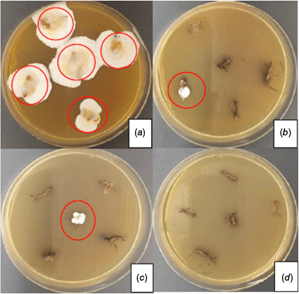
In collaborative research with the Brisbane City Council a Trichoderma-Actinomycete consortium used at 1 Trichoderma: 2 actinomycetes w/w ratio revealed improved biocontrol powers against P. noxium in the greenhouse and field applications.8–10 Trichoderma strains (#5001 and #5029) and actinomycetes (USC-595B and USC-6914) by themselves displayed -static activity rather than a -cidal one and the pathogen was able to recover (Fig. 2). Incorporated inoculum composed of Trichoderma strains and actinomycetes together showed strongest antagonist activity against P. noxium and the pathogen was not recovered (Fig. 2).
Recovery of Pyrrhoderma noxium from root samples (at the end of the experiment) as highlighted with red circles on Pyrrhoderma noxium selective media from the treatments of (a) Pyrrhoderma noxium; (b) Trichoderma consortium and Pyrrhoderma noxium; (c) actinomycetes consortium and Pyrrhoderma noxium; (d) Trichoderma and actinomycete consortium and Pyrrhoderma noxium (©H. Panchalingam). Red circles show Pyrrhoderma noxium survival in the port trial and subsequent growth on the agar plate.
In greenhouse trials initial screening (after 2 weeks of inoculation) of the BCAs and P. noxium confirmed the establishment of the inocula in the potting mix. During the experiment period none of the plants died due to P. noxium infection. However, the pathogen significantly reduced the plant growth (Fig. 3).
Effect of different treatments on plant growth parameters, (a) control; (b) P. noxium consortium; (c) Trichoderma consortium; (d) actinomycetes consortium; (e) Trichoderma consortium and actinomycetes consortium; (f) P. noxium with Trichoderma consortium; (g) P. noxium with actinomycetes consortium; (h) P. noxium with Trichoderma and actinomycete consortium) (©H. Panchalingam). Results were recorded after 6 months of pot trial.
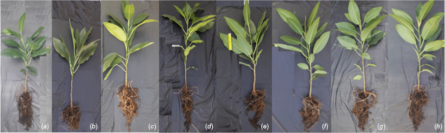
The effect of treatments on the growth of the plants used in the pot experiment was analysed using one-way ANOVA followed by Tukey’s test using SPSS statistics.9 Results indicated that Trichoderma and actinomycetes used in this study were effective in managing the pathogen. Moreover, significantly increased biological control activity was observed against the pathogen in the combined inoculum application rather than single biocontrol agent treatments (Trichoderma or actinomycetes).
The plant growth promotion observed in this study also indicated that the BCAs used in this study were not harmful to the plant, while P. noxium negatively impacted the plant growth (Fig. 3). Overall, results indicate that when supported by actinomycetes Trichoderma displays improved biological control and can be used against P. noxium to manage Brown root rot disease in public places and minimise the use of toxic chemical applications.
Data availability
The data that support this study are available in the article. For in-depth data, readers can refer to the Dissertations by Panchalingam42 and Adra.43
Acknowledgements
The authors gratefully acknowledge Mr Keith Foster (former Principal Arborist of the BCC) and the BCC for the funding provided. The authors thank Louisa Parkinson and Elizabeth Dann, QAAFI, UQ for providing P. noxium and Trichoderma strains used in the study as part of their collaboration with the BCC. Authors also thank Mr Richard Frenken (Director of Frenkenscapes Pty Ltd) and Dr Vatsal Naik for their assistance in the pot and field trials.
References
1 Chung CL, Lee TJ, Akiba M, Lee HH, Kuo TH, Liu D, Ke HM, Yokoi T, Roa MB, Lu MJ, Chang YY, Ann PJ, Tsai JN, Chen CY, Tzean SS, Ota Y, Hattori T, Sahashi N, Liou RF, Kikuchi T, Tsai IJ. Comparative and population genomic landscape of Phellinus noxius: a hypervariable fungus causing root rot in trees. Mol Ecol 2017; 26: 6301-6316.
| Crossref | Google Scholar | PubMed |
2 Chung CL, Huang SY, Huang YC, Tzean SS, Ann PJ, Tsai JN, Yang CC, Lee HH, Huang TW, Huang HY, Chang TT, Lee HL, Liou RF. The genetic structure of Phellinus noxius and dissemination pattern of brown root rot disease in Taiwan. PLoS One 2015; 10: e0139445.
| Crossref | Google Scholar | PubMed |
3 Hsiao WW, Hung TH, Sun EJ. The pathogenicity of basidiospores of Phellinus noxius which causes brown root rot disease in Taiwan. Taiwania 2019; 64: 263-268.
| Crossref | Google Scholar |
4 Sahashi N, Akiba M, Takemoto S, Yokoi T, Ota Y, Kanzaki N. Phellinus noxius causes brown root rot on four important conifer species in Japan. Eur J Plant Pathol 2014; 140: 869-873.
| Crossref | Google Scholar |
5 Farid MA, Lee SS, Maziah Z, Patahayah M. Pathogenicity of Rigidoporus microporus and Phellinus noxius against four major plantation tree species in Peninsular Malaysia. J Trop Sci 2009; 21(4): 289-298.
| Google Scholar |
6 Hodges CS, Tenorio JA. Root disease of Delonix regia and associated tree species in the Mariana Islands caused by Phellinus noxius. Plant Dis 1984; 68: 334-336.
| Crossref | Google Scholar |
7 Bolland L. Phellinus noxius: cause of a significant root-rot in Queensland hoop pine plantations. Aust For 1984; 47: 2-10.
| Crossref | Google Scholar |
8 Panchalingam H, Powell D, Adra C, Foster K, Tomlin R, Quigley BL, Nyari S, Hayes RA, Shapcott A, Kurtböke Dİ. Assessing the various antagonistic mechanisms of Trichoderma strains against the brown root rot pathogen Pyrrhoderma noxium infecting heritage fig trees. J Fungi (Basel) 2022; 8: 1105.
| Crossref | Google Scholar | PubMed |
9 Panchalingam H, Ashfield-Crook N, Naik V, Frenken R, Foster K, Tomlin R, Shapcott A, Kurtböke Dİ. Testing the Biocontrol Ability of a Trichoderma-Streptomycetes Consortium against Pyrrhoderma noxium (Corner) LW Zhou and YC Dai in Soil. J Fungi (Basel, Switzerland) 2022; 9: 67.
| Crossref | Google Scholar | PubMed |
10 Adra C, Panchalingam H, İpek Kurtböke D. Termite–gut-associated actinomycetes as biological control agents against phytopathogen Pyrrhoderma noxium. Microbiol Aust 2022; 43: 190-193.
| Crossref | Google Scholar |
11 Zhou LW, Ji XH, Vlasák J, Dai YC. Taxonomy and phylogeny of Pyrrhoderma: a redefinition, the segregation of Fulvoderma, gen. nov., and identifying four new species. Mycologia 2018; 110: 872-889.
| Crossref | Google Scholar | PubMed |
12 EFSA Panel on Plant Health (PLH), Bragard C, Baptista P, Chatzivassiliou E, Di Serio F, Gonthier P, Jaques Miret JA, Justesen AF, MacLeod A, Magnusson CS, Milonas P, Navas-Cortes JA, Parnell S, Potting R, Stefani E, Thulke HH, Van der Werf W, Vicent Civera A, Yuen J, Zappalà L, Golic D, Gobbi A, Maiorano A, Pautasso M, Reignault PL. Pest categorisation of Pyrrhoderma noxium. EFSA J 2024; 22(3): e8667.
| Crossref | Google Scholar | PubMed |
13 Garfinkel AR, Cannon PG, Klopfenstein NB, Stewart JE, Kim M-S. Identification of genetic groups within the invasive brown root rot pathogen, Pyrrhoderma noxium (formerly Phellinus noxius). In Reynolds GJ, Wilhelmi NP, Palacios compilers P, editors. Proceeding of the 66th Western International Forest Disease Work Conference; 3–7 June 2019; Estes Park, CO, WIFDWC; 2020. pp. 141–145.
14 Stewart JE, Kim MS, Ota Y, Sahashi N, Hanna JW, Akiba M, Ata JP, Atibalentja N, Brooks F, Chung C-L, Dann EK, Mohd Farid A, Hattori T, Lee SS, Otto K, Pegg GS, Schlub RL, Shuey LS, Tang AMC, Tsai J-N, Cannon PG, Klopfenstein NB. Phylogenetic and population genetic analyses reveal three distinct lineages of the invasive brown root-rot pathogen, Phellinus noxius, and bioclimatic modelling predicts differences in associated climate niches. Eur J Plant Pathol 2020; 156: 751-766.
| Crossref | Google Scholar |
15 Farid AM, Lee SS, Rosli HM, Maziah Z, Norwati M. Basal Root Rot, a new Disease of Teak (Tectona grandis) in Malaysia caused by Phellinus noxius. Malays J Microbiol 2005; 1: 40-45.
| Crossref | Google Scholar |
16 Ann PJ, Chang TT, Ko WH. Phellinus noxius brown root rot of fruit and ornamental trees in Taiwan. Plant Dis 2002; 86: 820-826.
| Crossref | Google Scholar | PubMed |
17 Schwarze FWMR, Jauss F, Spencer C, Hallam C, Schubert M. Evaluation of an antagonistic Trichoderma strain for reducing the rate of wood decomposition by the white rot fungus Phellinus noxius. Biol Control 2012; 61: 160-168.
| Crossref | Google Scholar |
18 Chang TT, Yang WW. Phellinus noxius in Taiwan: distribution, host plants and the pH and texture of the rhizosphere soils of infected hosts. Mycological Research 1998; 102(9): 1085-1088.
| Google Scholar |
19 Tchotet Tchoumi JM, Vivas M, Coetzee MPA, Rajchenberg M. Wood‐rotting basidiomycetes associated with declining native trees in timber‐harvesting compartments of the Garden Route National Park of South Africa. Austral Ecology 2017; 42: 947-963.
| Crossref | Google Scholar |
20 Lin CJ, Chung CH, Wu ML, Cho CL. Detection of Phellinus noxius decay in Sterculia foetida tree. J Trop For Sci 2013; 25(4): 487-496.
| Google Scholar |
21 Agustini L, Francis A, Glen M, Indrayadi H, Mohammed CL. Signs and identification of fungal root‐rot pathogens in tropical Eucalyptus pellita plantations. For Pathol 2014; 44: 486-495.
| Crossref | Google Scholar |
22 Mendgen K, Hahn M, Deising H. Morphogenesis and mechanisms of penetration by plant pathogenic fungi. Annu Rev Phytopathol 1996; 34: 367-386.
| Crossref | Google Scholar | PubMed |
23 Takashi M, Kikuno H, Kajikawa, A, Yokota, K. In planta distribution of Pyrrhoderma maxium, the causal agent of brown root rot of mango. AGRIS – International System for Agricultural Science and Technology, FAO; 2023. Available at https://agris.fao.org/search/en/providers/122430/records/66d56f89028a9120ab953fff
24 Chou H, Xiao YT, Tsai JN, Li TT, Wu HY, Liu LD, Tzeng DS, Chung CL. In Vitro and in Planta Evaluation of Trichoderma asperellum TA as a Biocontrol Agent Against Phellinus noxius, the Cause of Brown Root Rot Disease of Trees. Plant Dis 2019; 103: 2733-2741.
| Crossref | Google Scholar | PubMed |
25 Chang TT. Survival of Phellinus noxius in soil and in the roots of dead host plants. Phytopathology 1996; 86: 272-276.
| Crossref | Google Scholar |
26 Wu ZC, Chang YY, Lai QJ, Lin HA, Tzean SS, Liou RF, Tsai IJ, Chung CL. Soil is not a reservoir for Phellinus noxius. Phytopathology 2020; 110: 362-369.
| Crossref | Google Scholar | PubMed |
27 Godoy MG, Amorim GM, Barreto MS, Freire DMG (2018) Agricultural residues as animal feed: protein enrichment and detoxification using solid-state fermentation. In: Larroche C, Soccol CR, editors. Current Developments in Biotechnology and Bioengineering. Elsevier, Amsterdam; 2018. pp. 235–256. doi:10.1016/B978-0-444-63990-5.00012-8
28 Goodell B, Qian Y, Jellison J Fungal decay of wood: soft rot-brown rot-white rot. In: Schultz TP, Militz H, Freeman MH, Goodell B, Nicholas DD, editors. Development of Commercial Wood Preservatives: Efficacy, Environmental, and Health Issues, Vol. 982. Washington, DC: American Chemical Society; 2008. pp. 9–31. doi:10.1021/bk-2008-0982.ch002
29 Ibarra Caballero JR, Ata JP, Leddy KA, Glenn TC, Kieran TJ, Klopfenstein NB, Kim MS, Stewart JE. Genome comparison and transcriptome analysis of the invasive brown root rot pathogen, Phellinus noxius, from different geographic regions reveals potential enzymes associated with degradation of different wood substrates. Fungal Biol 2020; 124(2): 144-154.
| Crossref | Google Scholar | PubMed |
30 Dixon DJ. A taxonomic revision of the Australian Ficus species in the section Malvanthera (Ficus subg. Urostigma: Moraceae). Telopea 2003; 10: 125-153.
| Crossref | Google Scholar |
31 Truchan M, Tkachenko H, Buyun L, Kurhaluk L, Góralczyk A, Honcharenko V, Prokopiv A, Osadowski Z. Ficus Macrophylla Desf. ex Pers. Leaves (Moraceae) possess antimicrobial potential for the prevention of bacterial infections: preliminary in vitro study. Agrobiodiversity Improv Nutr, Health Life Qual 2019; 3: 117-121.
| Crossref | Google Scholar |
32 Tubby KV, Webber JF. Pests and diseases threatening urban trees under a changing climate. For Int J For Res 2010; 83: 451-459.
| Crossref | Google Scholar |
33 Rabiey M, Hailey LE, Roy SR, Grenz K, Al-Zadjali MAS, Glyn A. Barrett GA, Jackson RW. Endophytes vs tree pathogens and pests: can they be used as biological control agents to improve tree health? Eur J Plant Pathol 2019; 155: 711-729.
| Crossref | Google Scholar |
34 Moore T. Common solution to rotten problem in Brisbane parks. Brisbane Times, 25 May 2012. Available at https://www.brisbanetimes.com.au/national/queensland/common-solution-to-rotten-problem-in-brisbane-parks-20120524-1z6q4.html
35 Gezgin Y, Gül DM, Sözer Şenşatar S, Kara CU, Sargın S, Sukan FV, Eltem R. Evaluation of Trichoderma atroviride and Trichoderma citrinoviride growth profiles and their potentials as biocontrol agent and biofertilizer. Turk J Biochem 2019; 45: 1-13.
| Crossref | Google Scholar |
36 Sood M, Kapoor D, Kumar V, Sheteiwy MS, Ramakrishnan M, Landi M, Araniti F, Sharma A. Trichoderma: the “secrets” of a multitalented biocontrol agent. Plants (Basel, Switzerland) 2020; 9: 762.
| Crossref | Google Scholar | PubMed |
37 Benítez T, Rincón AM, Limón MC, Codón AC. Biocontrol mechanisms of Trichoderma strains. Int Microbiol 2004; 7: 249-260.
| Google Scholar | PubMed |
38 Kurtböke İ. Actinomycetes: what more can they offer in an era of metabolic engineering and artificial intelligence? Microbiol Aust 2025; 46(2): 72-76.
| Crossref | Google Scholar |
39 Kurtböke I. From Actinomycin onwards: Actinomycete success stories. Microbiol Aust 2012; 33(3): 108-110.
| Crossref | Google Scholar |
40 Yanti Y, Zainon MN, Umi Marshida AH. Antagonistic activity of three Actinomycetes, MG01, MG02 And KT2F towards Phellinus noxius. In: Business, Engineering and Industrial Applications (ISBEIA), IEEE Symposium, Bandung, Indonesia. 2012. pp. 729–732. doi:10.1109/ISBEIA.2012.6422986
41 Yaacob Y. Biocontrol of plant pathogen Phellinus noxius by selected actinomycetes, bacteria and fungi (MSc). Malaysia: Universiti Teknologi MARA; 2014. Available at https://ir.uitm.edu.my/id/eprint/16468/
42 Panchalingam H. Integrated management of Pyrrhoderma noxium by selected Trichoderma and actinomycete strains. PhD Dissertation, University of the Sunshine Coast, Queensland, Australia; 2022. doi:10.25907/00681
43 Adra C. The Biocontrol Potential of Termite-gut-associated Actinomycetes against Phytopathogenic Fungus Pyrrhoderma noxium. PhD Dissertation, University of the Sunshine Coast, Queensland, Australia; 2024. doi:10.25907/00819
Dr Harrchun Panchalingam completed his PhD under Dr Kurtböke’s supervision in 2022 at the University of the Sunshine Coast specialising in biological control methods against Pyrrhoderma noxium using Trichoderma species. Since then, he has been working as a lab supervisor and overses the production of Driscoll’s Australia berry tissue culture operation in Queensland. His research interests are biological control of plant diseases, development of biofertilisers, bioremediation of hydrocarbons and micropropagation of plants. |
Dr Cherrihan Adra is currently a Postdoctoral Research Fellow at the University of the Sunshine Coast (UniSC) within the Natural Product Chemistry research group. She was awarded her PhD in 2024 specialising in biological control methods against Pyrrhoderma noxium using natural products from termite-gut bacteria under Dr Kurtböke’s supervision. After completion of her PhD, she worked on UniSC’s Critical Zone Observation site where she applied Synchrotron-based techniques to investigate sulfur oxidation states in vadose zone soil. Currently, for her postdoctoral research, she is investigating the biological activities of natural products derived from stingless bee propolis including its antimicrobial, anti-inflammatory and anti-cancer properties. |
Prof. Alison Shapcott has been researching the population genetics and ecology of rainforest plants and palms since 1985. She is particularly interested in the evolution and maintenance of biodiversity. She has worked with rainforest types across Australia from Tasmania to Cape Tribulation and the Northern Territory as well as in Madagascar, Papua New Guinea, Djibouti and Brunei. She has established an extensive international network and regularly collaborates with researchers at the Royal Botanical Gardens of Kew (UK), the Smithsonian Institution NMNH (USA) and the Queensland Herbarium. |
Assoc. Prof. İpek Kurtböke has been working in the field of biodiscovery and has been an active member of the international actinomycete research community since 1982. She currently conducts research and teaches in the field of environmental and applied microbiology and biotechnology at the University of the Sunshine Coast, Queensland. She has also been an active member of the World Federation of Culture Collections (WFCC) and currently is the President of the Federation. She was also an Editorial Board member of Microbiology Australia for 20 years (2004–2024). |