

Postharvest Cercospora apii fruit rot disease on Cucurbita maxima (Cucurbitaceae)
M. Silva A and O. L. Pereira A BA Departamento de Fitopatologia, Universidade Federal de Viçosa, Viçosa, MG 36570-000, Brazil.
B Corresponding author. Email: oliparini@ufv.br
Australasian Plant Disease Notes 3(1) 21-23 https://doi.org/10.1071/DN08009
Submitted: 26 November 2007 Accepted: 10 March 2008 Published: 20 March 2008
Abstract
A new postharvest disease caused by Cercospora apii is reported for the first time on fruits of Cucurbita maxima.
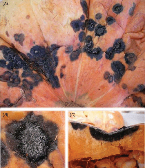
In June 2007, severe symptoms of necrotic rot were observed on fruits of Cucurbita maxima (Fig. 1), causing significant losses of ~4–5% at the market in Viçosa (MG, Brazil). According to the retailer, the fruits only showed disease symptoms ~7–10 days after putting them on the shelf. Structures of a cercosporoid fungus were associated with these necrotic fruit lesions.
|
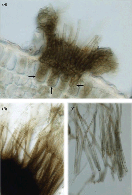
The fungus had the following morphology (VIC 30471): conidiophores arising from erumpent intraepidermal stromata, pigmented, pluriseptate, erect, straight, subcylindrical, strongly geniculate-sinuous, arranged in large dense sporodochial conidiomata. Conidiogenous cells integrated, terminal, with conspicuously thickened and darkened conidiogenous loci, mostly 2–3 µm wide. Conidia long, singly, acicular, hyaline, plurieuseptate, with truncate bases and subacute apices (Fig. 2). The fungus fitted well within the description of Cercospora apii s. lat. (Crous and Braun 2003).
|
The fungus was isolated from diseased fruits and mycelial plugs taken from a 20-day-old culture growing on V8 juice agar were utilised for the inoculation of healthy C. maxima fruits. The inoculated fruits were kept inside moistened plastic bags for 2 days at 28°C and then at 25°C for 14–15 days. Symptoms similar to those previously found were observed and the fungus was reisolated from the infected fruits. The control fruits, on which V8 juice agar plugs were deposited, remained healthy.
There are over 200 known cucurbit diseases of diverse aetiology. Zitter et al. (1998) listed Cercospora citrullina as the causal agent of cercospora leaf spot on cucurbits, later Crous and Braun (2003) listed C. citrullina as a synonym of C. apii. This species is known to cause leaf spots, typically only on the foliage, but they may appear on the petioles and stems if the environment is favourable for heavy sporulation. Cercospora apii has never been reported to cause lesions on fruits (Zitter et al. 1998) and this is, therefore, the first report of C. apii causing postharvest fruit rot disease on Cucurbita maxima. A possible quiescent mechanism will be investigated in a future study.