

Excision pathways for keratinocyte cancers diagnosed by teledermatology: a retrospective review
J. P. Tirado-Perez 1 * , A. Oakley 2 3 , R. Gansel 2
1
2
3
Abstract
The New Zealand population has one of the highest incidences of skin cancer in the world. Hospital waiting lists for surgical excision of keratinocytic skin cancers (basal cell carcinoma and squamous cell carcinoma) are lengthy, and increasingly, excisions are undertaken in primary care. Teledermatology, in response to general practitioners’ electronic referrals (e-referrals), can improve clinical communication between general practitioners and dermatologists.
The aim of this study was to evaluate an excision pathway for keratinocytic cancers diagnosed by teledermatology.
A retrospective observational descriptive review of a 3-month cohort of primary care e-referrals was undertaken.
Three hundred and fifty eight suspected keratinocytic cancers (KCs) were diagnosed by teledermatology; histology reports confirmed KC in 201 of 267 excisions (75%). The majority (77.2%) were excised by general practitioners an average of 25 days after the dermatologist’s recommendation. The rest were excised by plastic surgeons in private (3.4%) or at a public hospital (19.5%) after an average of 40 or 134 days, respectively.
E-referral pathways are now widely implemented. However, the ideal workflow for skin cancer management is unknown. We have demonstrated in New Zealand that surgery can be undertaken in primary care within a month of a teledermatology diagnosis and excision recommendation.
This study reports prompt excision of KCs by general practitioners after an e-referral and a teledermatology response.
Keywords: basal cell carcinoma, dermatologists, general practitioners, New Zealand, primary care, referrals, Skin cancer, squamous cell carcinoma, workflow.
| WHAT GAP THIS FILLS |
| What is already known: Keratinocytic cancers are increasing worldwide. Teledermatology is a valuable tool for communication between general practitioners, dermatologists, and plastic surgeons. |
| What this study adds: It describes the teledermatology diagnosis of 358 suspected keratinocytic cancers after e-referral, and pathways to excision by general practitioners, dermatologists, and plastic surgeons. |
Introduction
The ‘nonmelanoma’ skin cancers are predominantly keratinocytic cancers (KCs): basal cell carcinoma (BCC) and cutaneous squamous cell carcinoma (SCC). With an increasingly aged and sun-damaged population, excision of these tumours increases the burden on hospital surgical services. In Aotearoa New Zealand (NZ), more than 70 000 individuals were estimated to have been diagnosed with invasive KC in 2013, and 80 000 people were expected to be diagnosed in 2018 when the population was 4.9 million.1 More than one-third of patients with KC will develop at least one other KC over their lifetime.2 Diagnosis, treatment, and follow-up of KC are costly to the individual and public and private health services. The healthcare costs for new patients presenting with KCs were estimated to be NZ$ 129.4 in 2021.3
Several approaches to publicly funded skin cancer surgery in primary care have been initiated in NZ.4–7 E-referrals and teledermatology responses showed improved timeliness for melanoma surgery in primary care compared to hospital services in the Waikato.4 The Canterbury Initiative increased skin cancer surgery in primary care with a coordinated approach, including practical training, HealthPathways guidelines and e-referrals, and funding.5 A teledermoscopy service supports Waitematā’s e-referrals for suspected skin cancers, and many lesions are triaged to specialist-trained GPs for excision.6,7 Pathways to skin cancer diagnosis and management vary throughout NZ; Te Whata Ora does not employ dermatologists or plastic surgeons in all districts, and the training and surgical skills in primary care vary.
Teledermatology is also internationally recognised as an important form of healthcare delivery for diagnosing skin cancers.8,9
In the Waikato district, general practitioners (GPs) can refer patients with lesions suspicious of skin cancer to Dermatology for advice via a Suspected Skin Cancer pathway.4 During 2022, an average of 345 e-referrals were received each month for one to five specified lesions. Completing a lesion-specific template and attaching regional, close-up, and dermoscopic images are mandatory. The dermatologist responds with advice and treatment recommendations for each lesion. The main treatment for KC is complete surgical excision. 10 In NZ, GPs may undertake the surgery or refer to a plastic surgeon, dermatologist, or other specialist in a public hospital (where treatment for accepted patients is free) or in a private setting (insurance or self-funded).
This study’s primary objective was to determine the local excision pathways for KCs diagnosed by teledermatology after e-referral. The secondary objective was to determine how many suspected KCs were excised after a dermatological recommendation, by whom, and how long patients waited for their initial surgical procedure.
Methods
The study was registered with the local Clinical Audit Support Unit (4382P). It was a retrospective observational descriptive evaluation of primary care e-referrals received in one health district from 1 March 2022 to 31 May 2022 coded C449 (ICD-10 AM) by one of four teledermatologists. For each e-referral, we identified lesions diagnosed as KC and excluded non-KC.
We recorded demographic data (gender, age, ethnicity), the number of KCs per e-referral, clinical data (lesion location, referrer’s diagnosis, dermatologist’s diagnosis, and treatment recommendation), and the average response time. Using the hospital’s and private laboratory’s electronic records, we found the date of excision, histological report (histological diagnosis, completeness of excision and distance from the lateral and deep excision border to the tumour), and the identity of the surgeon.
Data were collected using a Microsoft Excel spreadsheet for descriptive statistical analysis. An ethics review is not required in NZ for a clinical audit.
Results
Demographic data (Table 1) were determined from all e-referrals coded as C449, which included 322 patients with an average age of 72.5 years (31–99); 45.03% were female with an average age of 73.7 years and 54.97% were male with an average age of 72.0 years. Ethnicity was most often recorded as NZ European (88.20%), other European (7.14%), or Māori (2.80%). The e-referrals recorded that 44.72% of patients had a history of skin cancer, more commonly in males (45.70% of males) than in females (43.50% of females). The average time for the dermatologist to respond to the referral was 13 days.
| N (322) | Frequency (%) | Age (mean, median, range) | ||
|---|---|---|---|---|
| Sex | ||||
| Male | 177 | 54.97 | 72.0, 74, 31–94 | |
| Female | 145 | 45.03 | 73.7, 74, 34–99 | |
| Ethnicity | ||||
| European – NZ | 284 | 88.20 | ||
| European – Other | 23 | 7.14 | ||
| Māori – NZ | 9 | 2.80 | ||
| European – not further defined | 3 | 0.93 | ||
| Latin American/Hispanic | 1 | 0.31 | ||
| African | 1 | 0.31 | ||
| Not stated | 1 | 0.31 | ||
| Skin cancer history | ||||
| No | 178 | 55.28 | ||
| Yes | 144 | 44.72 | ||
NZ, New Zealand.
Clinical data (Table 2) were determined for 358 lesions in 310 referrals after excluding incorrectly coded clinical diagnoses. Body location was selected using a drop-down menu, most often head and neck (36.59%), arm and hand (20.39%), or lower leg and foot (15.36%) and less often on the torso, back, thigh, and buttock. Lesions on the head and neck were most often on the nose, followed by the neck, mandibular area, forehead, ear, and scalp.
| N (358) | Frequency (%) | ||
|---|---|---|---|
| Location of lesion | |||
| Head and neck | 131 | 36.59 | |
| Arm and hand | 73 | 20.39 | |
| Lower leg foot | 55 | 15.36 | |
| Back | 43 | 12.01 | |
| Torso | 38 | 10.61 | |
| Thigh and buttock | 18 | 5.03 | |
| Referrer’s diagnosis | |||
| SCC | 165 | 46.09 | |
| BCC | 145 | 40.50 | |
| Unknown | 26 | 7.26 | |
| Seborrhoeic keratosis | 6 | 1.68 | |
| Other | 6 | 1.68 | |
| Melanocytic naevus | 5 | 1.40 | |
| Melanoma | 4 | 1.12 | |
| Bowen disease | 1 | 0.28 | |
| Dermatologist’s diagnosis | |||
| SCC | 165 | 46.09 | |
| BCC | 158 | 44.13 | |
| Keratinocytic skin cancer | 35 | 9.78 | |
| Dermatologist recommendation | |||
| Excision | 350 | 97.8 | |
| Topical treatment | 3 | 0.84 | |
| Biopsy or monitor | 2 | 0.56 | |
| Biopsy | 1 | 0.28 | |
| Cryosurgery or topical treatment | 1 | 0.28 | |
| Radiotherapy or monitor | 1 | 0.28 | |
SCC, squamous cell carcinoma; BCC, basal cell carcinoma.
The referrer diagnosis was most often SCC (46.09%) or BCC (40.50%). Other suspected diagnoses included seborrhoeic keratosis, melanoma, melanocytic naevus, SCC in situ, wart, unknown, or other. The dermatologist diagnosed SCC (46.09%), BCC (44.13%), or keratinocytic skin cancer (9.78%) when either SCC or BCC was likely. Diagnostic concordance between referrer and dermatologist was 73.9% for SCC and 67.0% for BCC.
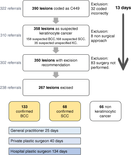
Excision was recommended for 350 lesions (97.8%) in 302 patients. Other treatment suggestions were to monitor, topical treatment (usually imiquimod), incisional biopsy, radiotherapy, and cryosurgery. The flowchart (Fig. 1) demonstrates the pathway and timeliness of the lesions. A total of 267 excisions were completed, while 83 excisions were not carried out (Supplementary Table S5). Most excisions were performed by GPs (77.15%) after an average of 25.2 days, excluding 20 excisions in primary care before receiving the dermatological recommendation. The rest were performed by the plastic surgical department (22.85%) in a public hospital (19.48%) or plastic surgeons in a private clinic (3.37%) after an average of 133.8 and 39.6 days, respectively. Excision specimens were analysed by a private laboratory (80.52%) or by the public hospital laboratory (19.48%) (Table 3).
Flowchart showing keratinocyte cancer excision pathways after teledermoscopy diagnosis. BCC, basal cell carcinoma; SCC, squamous cell carcinoma; KC, keratinocytic cancer.
| N (267) | Frequency (%) | Days to excision | ||
|---|---|---|---|---|
| Type of practice | ||||
| Primary care | 206 | 77.15 | 25.2 | |
| Hospital Department of Plastic Surgery | 52 | 19.48 | 133.8 | |
| Private plastic surgeon | 9 | 3.37 | 39.6 | |
| Laboratory | ||||
| Private | 215 | 80.52 | ||
| Hospital | 52 | 19.48 | ||
N, number of lesions.
Histology reports of the 267 excisional biopsies included 201 KCs (75.3%): 68 SCCs and 133 BCCs (Table 4). The teledermatologist diagnosis was concordant with histology in 181 lesions (67.8%), being higher for BCC (87.9%) than SCC (48.8%) (Table 4). The distance of the tumour from the nearest resection lateral and deep border in millimetres (mm) was recorded for confirmed KCs. Details of excision margins have been previously reported.11
| Dermatological and histological diagnosis | N (267) | Frequency (%) | Concordance (%) | |
|---|---|---|---|---|
| Dermatologist diagnosed SCC | 129 | 48.31 | ||
| SCC | 63 | 23.60 | 48.8 | |
| BCC | 17 | 6.37 | ||
| Solar keratosis | 15 | 5.62 | ||
| Seborrhoeic keratosis | 7 | 2.62 | ||
| In situ SCC | 7 | 2.62 | ||
| Epidermal inclusion cyst | 4 | 1.50 | ||
| Wart | 3 | 1.12 | ||
| Scar | 3 | 1.12 | ||
| Lichen planus-like keratosis | 2 | 0.75 | ||
| Dermatofibroma | 1 | 0.37 | ||
| Hyperkeratosis, chronic folliculitis | 1 | 0.37 | ||
| Hyperkeratosis, possible porokeratosis | 1 | 0.37 | ||
| Melanoma | 1 | 0.37 | ||
| Prurigo nodularis | 1 | 0.37 | ||
| Solar elastosis | 1 | 0.37 | ||
| Solar keratosis with lichen planus-like inflammation | 1 | 0.37 | ||
| Vascular malformation | 1 | 0.37 | ||
| Dermatologist diagnosed BCC | 116 | 43.45 | ||
| BCC | 102 | 38.20 | 87.9 | |
| SCC | 3 | 1.12 | ||
| Dermatofibroma | 2 | 0.75 | ||
| No evidence of malignancy | 2 | 0.75 | ||
| Dermal fibrosis | 1 | 0.37 | ||
| In situ SCC | 1 | 0.37 | ||
| In situ SCC and seborrhoeic keratosis | 1 | 0.37 | ||
| Scar | 1 | 0.37 | ||
| Seborrhoeic keratosis | 1 | 0.37 | ||
| Solar keratosis | 1 | 0.37 | ||
| Superficial leiomyosarcoma | 1 | 0.37 | ||
| Dermatologist diagnosed keratinocytic skin cancer | 22 | 8.24 | ||
| BCC | 14 | 5.24 | 72.7 | |
| Solar keratosis | 4 | 1.50 | ||
| SCC | 2 | 0.75 | 72.7 | |
| In situ SCC | 2 | 0.75 |
N, number of lesions; SCC, squamous cell carcinoma; BCC, basal cell carcinoma.
Discussion
We have described a successful e-referral pathway in one health district between GPs and teledermatologists for managing KCs. There was diagnostic concordance between the referrer and responding dermatologist in more than two-thirds of lesions, reflecting good knowledge and skills in primary care. GPs performed most of the excisions within a month of referral. In 20 cases, this was before the response had been received, which may have been due to perceived urgency or convenience.
There is growing interest in implementing teledermatology for the diagnosis of skin cancer.12 In many reports, teledermatology has shown a higher sensitivity for cancer detection than face-to-face examination, providing high-resolution images are received, including dermoscopy images.12 Our teledermatologists’ diagnostic uncertainty was mainly due to referrer nonadherence to the e-referral requirements for regional, close-up, and dermoscopy images of high resolution (>2000 × 1500 pixels), in focus, well-lit, and with a plain background.
Three-quarters of the lesions were excised in primary care, including some high-risk lesions on the head and neck. Patients waited an average of 108 days longer to have an excision undertaken at the public hospital than in primary care. Using GP skin surgeons to excise KCs mitigates the long waitlists for hospital surgical excision. Treatment in primary care may incur costs to the patient and a higher risk of the surgery being incomplete, with our study finding that 15 of 201 KCs were incompletely excised in primary care, necessitating further treatment.11 We recommend following the minimal clinical margin guidelines of 4 mm for SCC and 3 mm for BCC.13–15
An analysis of why surgery was not performed in 23.71% of lesions when recommended is out of scope for this study as we did not have access to the primary care records. Eight lesions were treated non-surgically in primary care despite the dermatologist recommending excision, and 20 lesions were excised before the recommendation was received.
Conclusion
We have described a collaborative skin cancer workflow in one health district in New Zealand using teledermatology responses to e-referrals made by general practitioners. Teledermatology is unavailable in many districts of NZ where there is no dermatologist or other expert in skin cancer diagnosis. Referrers to a teledermatology service should be provided with a template to remind them to include relevant patient risk factors and lesion characteristics. To optimise diagnostic quality, they should attach high-resolution, regional, close-up, and dermoscopy images. Excision is usually recommended for suspected KC. General practitioners play an essential role in carrying out most surgeries within a month of the recommendation. They should be trained, credentialed according to their skills, and follow surgical guidelines.
References
1 Sneyd MJ, Gray A. Expected non melanoma skin (Keratinocytic) cancer incidence in New Zealand for 2018. Wellington: Health Promotion Agency; 2018. Available at https://www.hpa.org.nz/sites/default/files/Expected%20Non%20Melanoma%20Skin%20KC%20incidence%20in%20NZ%20for%202018_FinalReport_777173.pdf
2 Stratigos AJ, Garbe C, Dessinioti C, et al. European interdisciplinary guideline on invasive squamous cell carcinoma of the skin: Part 1. epidemiology, diagnostics and prevention. Eur J Cancer 2020; 128: 60-82.
| Crossref | Google Scholar | PubMed |
3 Gordon LG, Shih S, Watts C, et al. The economics of skin cancer prevention with implications for Australia and New Zealand: where are we now? Public Health Res Pract 2022; 32(1): 31502119.
| Crossref | Google Scholar | PubMed |
4 Na H, Oakley A. Timeliness of diagnosis and treatment of cutaneous melanoma with dermatology, general practice, plastics surgery collaboration – are we meeting standards? J Prim Health Care 2023; 15: 267-73.
| Crossref | Google Scholar | PubMed |
5 McGeoch G, Sycamore M, Shand B, et al. A regional programme to improve skin cancer management. J Prim Health Care 2015; 7: 339-44.
| Crossref | Google Scholar | PubMed |
6 Sunderland M, Teague R, Gale K, et al. E-referrals and teledermatoscopy grading for melanoma: a successful model of care. Australas J Dermatol 2020; 61(2): 147-51 Epub 16 February 2020.
| Crossref | Google Scholar | PubMed |
7 Wen D, Gale K, Martin R. Quality assessment of a large primary GP skin cancer service in Auckland, New Zealand. N Z Med J 2020; 133(1509): 17-27.
| Google Scholar | PubMed |
8 Marwaha SS, Fevrier H, Alexeeff S, et al. Comparative effectiveness study of face-to-face and teledermatology workflows for diagnosing skin cancer. J Am Acad Dermatol 2019; 81(5): 1099-106.
| Crossref | Google Scholar | PubMed |
9 Ferrandiz L, Moreno-Ramirez D, Nieto-Garcia A, et al. Teledermatology-based presurgical management for nonmelanoma skin cancer: a pilot study. Dermatol Surg 2007; 33(9): 1092-8.
| Crossref | Google Scholar | PubMed |
10 Nolan GS, Kiely AL, Totty JP, et al. Incomplete surgical excision of keratinocyte skin cancers: a systematic review and meta‐analysis. Br J Dermatol 2021; 184: 1033-44.
| Crossref | Google Scholar | PubMed |
11 Tirado-Pérez J-P, Oakley A. Surgical excision margins in primary care and plastic surgery for keratinocytic cancers diagnosed via teledermatology: retrospective observational cross-sectional study. iproc 2023; 9: e49466.
| Crossref | Google Scholar |
12 Jones LK, Oakley A. Store-and-forward teledermatology for assessing skin cancer in 2023: literature review. JMIR Dermatol 2023; 6: e43395.
| Crossref | Google Scholar | PubMed |
13 Kim JYS, Kozlow JH, Mittal B, et al. Guidelines of care for the management of cutaneous squamous cell carcinoma. J Am Acad Dermatol 2018; 78(3): 560-78.
| Crossref | Google Scholar |
14 Stratigos AJ, Garbe C, Dessinioti C, et al. European interdisciplinary guideline on invasive squamous cell carcinoma of the skin: Part 2. Treatment. Eur J Cancer 2020; 128: 83-102.
| Crossref | Google Scholar | PubMed |
15 Peris K, Fargnoli MC, Garbe C, et al. Diagnosis and treatment of basal cell carcinoma: European consensus–based interdisciplinary guidelines. Eur J Cancer 2019; 118: 10-34.
| Crossref | Google Scholar | PubMed |