

Mapping the trajectory of acute mild-stroke cognitive recovery using serial computerised cognitive assessment
Alana Campbell A B * , Louise Gustafsson C , Rohan Grimley B D , Hannah Gullo A , Ingrid Rosbergen B E and Mathew Summers F
A
B
C
D
E
F
Abstract
Cognitive impairment is common post-stroke. There is a need to understand patterns of early cognitive recovery post-stroke to guide both clinical and research practice. The aim of the study was to map the trajectory of cognitive recovery during the first week to 90-days post-stroke using serial computerised assessment.
An observational cohort study recruited consecutive stroke patients admitted to a stroke unit within 48 hours of onset. Cognitive function was assessed using the computerised Cambridge Neuropsychological Test Automated Battery (CANTAB) daily for seven days, then 14, 30 and 90 days post-stroke. The CANTAB measured visual episodic memory and learning, information processing speed, visuo-spatial working memory, complex sustained attention and mental flexibility. Repeated measures MANOVA/ANOVA with Least Squares Difference post-hoc analyses were performed to ascertain significant change over time.
Forty-eight participants, mean age 73, primarily mild, ischaemic stroke, completed all assessment timepoints. There was a trajectory of early, global cognitive improvement, indicative of a post-stroke delirium, that largely stabilised between 6 and 14-days post-stroke. Change over time was examined within each cognitive test, with one measure stabilising by day 6 (Reaction Time) and others detecting improving performances up to 14 days post-stroke.
Serial, computerised cognitive assessment can effectively map post-stroke cognitive recovery and revealed an early phase of global improvement over 14 days that is evidence for an acute post-stroke delirium. Resolution of post-stroke delirium in the second week following mild stroke indicates more extensive neuropsychological testing may be undertaken earlier than previously thought.
Keywords: Cognitive dysfunction, stroke, delirium, mild cognitive impairment, neuropsychological tests.
Introduction
Stroke continues to be the second leading cause of death and disability globally (Krishnamurthi et al., 2020), with 30-40% of stroke-survivors experiencing persistent cognitive deficits (McDonald et al., 2019). Post-stroke cognitive impairment impacts individual quality of life (Barker-Collo et al., 2010; Nys et al., 2006) and function (Tatemichi et al., 1994) and increases burden on communities (Krishnamurthi et al., 2020). There is a need to understand cognitive recovery patterns post-stroke to compare efficacy of therapeutic interventions against the rate of natural post-stroke improvement (Duncan et al., 2000). A growing body of literature has described the long-term trajectory of post-stroke cognitive change (Levine et al., 2015; Lo et al., 2021; Mahon et al., 2017; Zheng et al., 2019) with general consensus that early cognitive deficits are complex and multifactorial due to processes of neuro-vascular injury and recovery, delirium, fatigue, fluctuating alertness and emotional distress (Lezak, 2012; McDonald et al., 2019). However, there is limited information on the trajectory of cognitive recovery over the first week post-stroke, when cognition is impacted by the acute effects of stroke, despite the clinical requirement of early assessment to support critical decisions such as discharge disposition and referral to rehabilitation services (Australian Commission on Safety & Quality in Health Care, 2019; Lindsay et al., 2014).
Cognitive screening post mild-stroke is essential as cognitive impairment is pervasive (Boulanger et al., 2018; Lanctôt et al., 2020; Pendlebury et al., 2015; Stroke Foundation, 2022) and can result in reduced participation (Adamit et al., 2014; Tellier & Rochette, 2015). With short length-of-stay in acute care, screening measures are conducted expeditiously post-stroke (Bertolin et al., 2018; Verdelho et al., 2021). Brief screening and more extensive neuropsychological testing are feasible early post-stroke (Nys et al., 2005; van Zandvoort et al., 2005), with screening tests more efficient and better tolerated by patients in acute care. However, there is no gold standard for cognitive screening (Hachinski et al., 2006; Quinn et al., 2018; Quinn et al., 2021), with measures needing to cover a broad range of cognitive domains, be valid, feasible and sensitive for identification of impairments post-stroke (Chan et al., 2017; Chan et al., 2014; Stolwyk et al., 2014). They also only provide a ‘snapshot’ at a single point in time and should be responsive to change for monitoring of cognitive recovery (Skirrow et al., 2021), making it difficult for one tool to meet all requirements. Exploring new technologies could provide alternatives for measuring cognition in acute stroke.
Computerised cognitive assessments offer sensitive continuous measures that can be customised for select subtests and repeated to mark changes over short epochs of cognitive recovery (Aslam et al., 2018; Pettigrew et al., 2021; Zygouris & Tsolaki, 2015), such as the acute phase post-stroke (Bernhardt et al., 2017). Computerised cognitive assessment platforms are feasible as research measures acutely post-stroke (Cumming et al., 2012; Shopin et al., 2013) and are designed for serial measurement of cognition over short time intervals (Cambridge Campos-Magdaleno et al., 2021; Cognition, 2022; Skirrow et al., 2021), but have not been used in both capacities in the acute post-stroke period. We aimed to map the trajectory of cognitive recovery during the first week post-stroke and up to 90-day follow-up using serial computerised cognitive assessment.
Materials and methods
Patients and setting
A prospective, single-centre, observational, cohort study was conducted in the Acute Stroke Unit of the Sunshine Coast University Hospital. Ethical approval was obtained from: The Prince Charles Hospital Human Research Ethics Committee, HREC/17/QPCH/163; the University of Queensland (#2017001149); and from University of the Sunshine Coast (S/17/1091). Data were collected from January to September 2018.
Participant screening included all patients diagnosed with stroke by senior medical consultants. Eligible patients were aged 18 years or older and within 48 hours of stroke onset. Patients were excluded if they: were palliative in treatment intent; previously had diagnoses of dementia or extensive mental illness; were blind or were anticipated to be unavailable for follow-up. The Principal Investigator provided study information to all consecutive eligible patients and invited them to participate. Signed consent was obtained from participants or substitute decision-makers.
Data collection tools
Demographic information - age, gender, premorbid function, education, occupation and comorbid medical conditions - were gathered from medical records and participant interview. Admission medical entries and imaging (Computerised Tomography, CT and Magnetic Resonance Imaging, MRI) provided stroke-specific information: National Institutes of Health Stroke Scale (NIHSS) (Brott et al., 1989) for stroke severity (Adams et al., 1999), stroke type, hemisphere, symptoms and reperfusion therapies.
Participants were screened for cognitive confounders: the Depression, Anxiety and Stress Scale-21 assessed for clinical symptoms of anxiety and depression (Ng et al., 2007) and was provided to the patient to read while questions were read through with the primary investigator; the Wechsler Test for Adult Reading (WTAR) estimated pre-stroke intellectual capacity (Full Scale intelligence Quotient, FSIQ) (Steward et al., 2016) and the 4-A’s Test screened for delirium (4-AT) (Lees et al., 2013).
Data were collected from Version 7.1 of the Montreal Cognitive Assessment (MoCA); routinely used by local clinicians to screen cognitive function for assessment of rehabilitation needs post-stroke. The MoCA screens cognitive domains of Executive Function, Naming, Attention, Language, Abstraction, Delayed Recall and Orientation (Nasreddine et al., 2005). It has been shown to predict long-term cognitive and functional outcomes, and is valid and feasible in the acute phase post-stroke (Abzhandadze et al., 2019; Chiti & Pantoni, 2014; Koski, 2013; Zietemann et al., 2018).
Cambridge neuropsychological test automated battery (CANTAB)
The CANTAB (Connect version) was the primary cognitive outcome measure. The CANTAB is a computerised, portable (tablet-based), cognitive assessment battery that presents parallel versions and randomised stimuli to minimise practice effects for extensive serial assessment (Cambridge Cognition, 2021). Automated scoring and instruction remove human error, reduce variance in administration and allow non-specialist staff to administer tests. Additionally, on reaction time-based measures, time to move is partitioned from cognitive processing time to decide and react, enabling the separation of cognitive speed from impaired motor functions post-stroke. The subtests primarily utilise visual stimuli, reducing language dependent effects and allowing those with language impairment to participate in testing (Cacciamani et al., 2018; Crivelli et al., 2021). Additionally, the CANTAB platform uses embedded practice sessions to ensure participants can successfully perform the task before proceeding to the assessment phase of the test.
The CANTAB features 23 modular subtests, with studies on subtest properties and availability of normative data confined to a limited set (Aslam et al., 2018). Subtest selection was determined by domains of post-stroke cognitive impairment identified in previous research (Cumming et al., 2013; Mole & Demeyere, 2020) and limited by overall time to perform the customised battery, accommodating for participants’ reduced tolerance early post-stroke. Verbal measures were not selected, for inclusivity of people with aphasia and as aphasia is routinely screened separately. The selected CANTAB measures are outlined in Table 1, in order of presentation for testing.
CANTAB Subtests and Key Measures
| Subtest | Key Measures | |
|---|---|---|
| Paired Associates Learning (PAL) A measure of visual episodic memory and learning Patterns are revealed in boxes on the screen (adjustingto different levels of up to 8 patterns to avoid ceilingeffects) and participants must remember where eachpattern was located. | PAL Total Errors Adjusted (PALTEA) Raw score total number of errors adjusted for incomplete trials. The greater the number of errors the worse the memory performance. This measure allows comparison on performance to be made acrossall participants regardless of those that were unable to complete the finalstage of the task. | |
| Five-Choice Reaction Time (RTI) A measure of information processing speed and attention. Participants hold their finger on a graphic ‘button’ untilone of five circles flashes yellow. As swiftly as possible, theparticipant must touch the circle which flashed and thenreturn their finger to the button. | RTI Mean 5-Choice Reaction Time (RTIFMRT) Average reaction time (msec) on the 5 choice RTI task excluding time takento move to the target. Mean RT is a relatively pure measure of attentionalspeed and resources. RTI Mean 5-Choice Movement Time (RTIFMMT) Average movement time (msec) in responding to the 5 choice RTI tasks.This is a measure of motor response speed independent of attentionalresources. | |
| Spatial Working Memory (SWM) A measure of visual working memory capacity. A task where participants try to locate a yellow squareconcealed in an array of red boxes but must recall wherethey have previously located a yellow square and avoidthose boxes. | SWM Total Errors (SWMTE) Raw score total number of errors adjusted for incomplete trials. Greaterthe number of errors the worse the working memory capacity. SWM Strategy (SWMS) Raw strategy score assessing strategy use by participant across trials.This measure uses a composite score of various forms of errors made inperforming the task. | |
| Rapid Visual Processing (RVP) A measure of signal detection sensitivity and sustained attention. Participants are required to rapidly process a string of targetsequences with digits presented at a rate of 100 digits/minute. | Rapid Visual Processing ‘A’ Prime (RVP A’) Calculates detection of the target sequence and is a precise measure ofattentional processing capacity. RVP A’ is a ratio score, with higher scoresindicating improved attentional capacity. | |
| Multi-tasking Test (MTT) A measure of executive function through mental flexibility tasksinvolving random rule shifts, as well as congruency with symbolmeaning vs physical location. | MTT Incongruent Trial Mean Latency (MTTLNOM) Calculates the mean latency in decision time (msec) on the conflictualincongruent trial. |
Procedure
Following enrolment, participant demographic and stroke-specific information were collected. CANTAB assessment was performed daily for 7 days, with follow-up at 14, 30 and 90-days post-stroke (+3 days for 30 and 90-day follow-up) in hospital and subsequent community settings post-discharge. The MoCA, version 7.1 was administered within the first week as per standard clinical practice. The 4-AT was conducted from the date of enrolment, and the DASS-21and WTAR were administered in the first four days with DASS-21 repeated at 14 days. For participants exhibiting symptoms of delirium (scoring ≥4), the 4-AT was repeated, and the number of days with clinically recognised delirium was recorded.
Statistical analysis
Analyses were performed using IBM SPSS for Windows (version 27.0). Participants with missing data for all CANTAB subtests in a single assessment timepoint were removed from analysis (incomplete data). Participants with missing data for some (not all) CANTAB subtests within a timepoint or over multiple timepoints were retained.
Independent samples t-test and Chi-Square were used to compare participants who completed all timepoints with those who did not across key demographic variables. Variables from similar cognitive domains were entered into a repeated measures MANOVA to control for study-wide type I error rate. Significant multivariate tests were followed by repeated measures ANOVAs for each of the variables corrected for family-wise error, to determine significant change over time. Unrelated variables were analysed using independent repeated measures ANOVAs. Post-hoc analyses of change over time were examined by Least Square Differences comparison (p < .05).
Results
Participant information
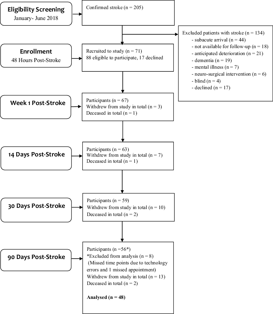
Of the 88 patients determined eligible to participate, 17 declined and 71 participants were recruited. Thirteen of the 71 participants withdrew prior to 90-day follow-up: four were not accessible for follow-up, two had ongoing medical issues, and 7 declined to participate further. Two participants died, seven missed an entire assessment time point due to technology errors and one failed to attend an appointment. A final sample of 48 participants was available for trajectory analysis (Fig. 1).
Demographic data
The 48 participants were equally distributed for gender and stroke hemisphere, with a mean age of 73 years, and the majority sustained a mild, ischaemic stroke (NIHSS <6). Average length of stay in acute care approached 5 days with 25% of participants exhibiting symptoms of delirium. More than 40% exhibited symptoms of depression the first week, dropping to 30% by day 14. As per usual clinical practice, participants with functional language deficits were screened using the Western Aphasia Battery. Three participants had moderate and three mild aphasia, with one omitted from analysis due to technology error. Three of these participants could perform the WTAR at 14 days. One was also unable to complete DASS-21 or MoCA the first week but could participate at day 14. All could perform the CANTAB. MoCA mean score was 22.7 with over half scoring at or above 23 (Table 2).
Demographic and Clinical Characteristics of Participants with Data for All Timepoints vs Participants with Missing Data in One or More Timepoints Post-stroke (n = 71)
| n (%) or mean (+SD) | Participants with no missing time points | Participants with missing time points | Comparison of participant groups | |||
|---|---|---|---|---|---|---|
| (n = 48) | (n = 23) | P < 0.05†† (95% CI) | ||||
| Sex (Female) | 23 (48%) | 7 (30%) | .163 | |||
| Age (years) § | ||||||
| Mean (SD) | 72.7 (10.4) | 78.2 (11.8) | .353 | |||
| Median | 72.5 | 80.5 | ||||
| Range | 51–91 | 44–96 | ||||
| Education (years) ‖ | ||||||
| Mean (SD) | 11.3 (2.6) | 12 (3.3) | .195 | |||
| Median | 10.5 | 12 | ||||
| Range | 7–19 | 4–18 | ||||
| Stroke type | ||||||
| Ischaemic | 45 (94%) | 22 (96%) | —— | |||
| Haemorrhagic | 3 (6%) | 1 (4%) | —— | |||
| Hemisphere | ||||||
| L | 23 (48%) | 8 (35%) | —— | |||
| R | 21 (44%) | 13 (56%) | —— | |||
| Bilateral | 4 (8%) | 2 (9%) | —— | |||
| Severity | ||||||
| Mild | 46 (96%) | 17 (74%) | .005* | |||
| Mod | 2 (4%) | 5 (22%) | ||||
| Severe | 0 | 1 (4%) | ||||
| Aphasia on admission | 5 (10%) | 1 (4%) | —— | |||
| LOS acute (days) | ||||||
| Mean (SD) | 4.8 (2.4) | 9.9 (8.4) | .009* | |||
| Median | 4.5 | 8 | ||||
| Range | 1–11 | 2–34 | ||||
| MoCA | ||||||
| Completed | 46 (96%) | 16 (70%) | —— | |||
| Mean (SD) | 22.7 (4.3) | 22.6 (3.5) | .888 | |||
| Range | 12–30 | 18–29 | ||||
| Score ≥23 | 27 (56%) | 8 (35%) | .546 | |||
| Participants with delirium | 12 (25%) | 10 (44%) | .087 | |||
| Days of delirium | ||||||
| Mean (SD) | 0.7 (1.4) | 4.2 (8.7) | .064 | |||
| Median | 0 | 0 | ||||
| Range | 0–5 | 0–34 | ||||
| WTAR (Estimated FSIQ) | ||||||
| Mean (SD) | 103.26 (8.4) | 106.1 (7.5) | .514 | |||
| Median | 101 | 106.1 | ||||
| Range | 84–117 | 91–118 | ||||
| Depression | Day 4 | Day 14 | Day 4 | Day 14 | ||
| n = 48 | n = 48 | n = 21 | n = 16 | |||
| Normal | 27 (56%) | 33 (69%) | 13 (62%) | 12 (75%) | 0.417 Day 4 | |
| Mild | 6 (13%) | 4 (8%) | 1 (5%) | 0 (0%) | ||
| Moderate | 10 (21%) | 7 (15%) | 2 (10%) | 1 (6%) | ||
| Severe | + 4 (8%) | 4 (8%) | 2 (10%) | 1 (6%) | ||
| Unable | 1 (2%) | 0 (0%) | 2 (10%) | 2 (13%) | ||
| Anxiety | ||||||
| Normal | 31 (65%) | 31 (65%) | 15 (71%) | 11 (69%) | 0.262 Day 4 | |
| Mild | 5 (10%) | 5 (10%) | 1 (5%) | 2 (13%) | ||
| Moderate | 6 (13%) | 4 (8%) | 1 (5%) | 1 (6%) | ||
| Severe | + 5 (10%) | 8 (17%) | 1 (5%) | 0 (0%) | ||
| Unable | 1 (2%) | 0 (0%) | 2 (10%) | 2 (13%) | ||
| Stress | ||||||
| Normal | 34 (71%) | 36 (75%) | 14 (67%) | 10 (63%) | —— | |
| Mild | 6 (13%) | 1 (2%) | 1 (5%) | 3 (19%) | ||
| Moderate | 6 (13%) | 7 (15%) | 2 (10%) | 0 (0%) | ||
| Severe | + 1 (2%) | 3 (6%) | 1 (5%) | 1 (6%) | ||
| Unable | 1 (2%) | 0 (0%) | 2 (10%) | 2 (13%) | ||
†Data p values determined by Chi-square for categorical variables (Gender, Delirium, Stroke Hemisphere, Stroke Severity, Stroke Type, Depression, Anxiety, Stress) and Independent-Samples t-tests, for the remainder of the comparisons.
†Where cell is blank, insufficient n for analysis.
§21 year-old outlier removed.
‖Academic professor with multiple PhD’s removed as outlier.
¶Ischaemic includes stroke w/ haemorrhagic transformation.
The only significant demographic differences between participants analysed and those excluded from analysis was acute care length of stay which was nearly twice as long for those excluded. There was a trend toward greater rates of delirium in excluded participants, however mean MoCA scores were similar between groups suggesting attention difficulties associated with delirium were not severe enough to adversely impact a cognitive screening measure. Three participants in the excluded group had delirium for >10 days. For included participants, delirium clinically resolved an average of 4 days post-stroke. Lastly, a significantly higher proportion of participants with moderate to severe stroke had incomplete data (74%) compared to mild stroke (26%) (Table 2).
Trajectory data
Raw mean scores are summarised in Table 3. Standard scores were available for PALTEA and RVP A’, which indicate that participants’ average scores were trending below a healthy adult population at all time-points for these subtests.
CANTAB Subtest Mean Scores by Day Post-stroke
| CANTAB Subtests† | PALTEA Visual episodic memory and learning total errors adjusted | RTIFMRT Reaction time (mS) | RTIFMMT Movement time (mS) | SWMTE Spatial working memory total errors | SWMS Spatial working memorystrategy use | RVP A′ Visual sustained attention sensitivity to target sequence | MTTLNOM Multitasking test mean latency of response (mS)mental flexibility | ||||||||||||
|---|---|---|---|---|---|---|---|---|---|---|---|---|---|---|---|---|---|---|---|
| n | X̅ (SE) Range | z- Score X̅ (SE) Range | %ile Score X̅ | n | X̅ (SE) Range | n | X̅ (SE) Range | n | X̅ (SE) Range | n | X̅(SE) Range | n | X̅ (SE) Range | z- Score X̅ (SE) Range | %ile Score X̅ | n | X̅ (SE) Range | ||
| Day 2 | 48 | 43.7(2.6) 7.0-68.0 | −1.23(0.13) −2.3 to 0.44 | 19 | 45 | 525.7(22.0) 362-1309 | 45 | 415.8(21.1) 201-820 | 45 | 21.2(1.1) 0.00-39.00 | 45 | 9.24(0.3) 2.0-12.0 | 42 | 0.82(0.01) 0.68-0.92 | −1.14(.08) −2.3-0.03 | 17 | 41 | 1001.0(16.5) 748-1216 | |
| Day 3 | 48 | 37.9*(2.6) 8.0-69.0 | −0.94(0.13) −2.3 to 0.64 | 25 | 48 | 516.9(18.9) 348-1048 | 48 | 409.8(26.8) 202-1215 | 48 | 21.0(1.4) 0.00-47.00 | 48 | 9.40(0.3) 3.0-13.0 | 45 | 0.83(0.01) 0.67-0.94 | −0.98(.10) −2.3-0.58 | 21 | 44 | 998.8(21.3) 724-1356 | |
| Day 4 | 47 | 36.6(2.8) 5.0–67.0 | −0.84(0.14) −2.3 to 0.99 | 28 | 47 | 486.4(18.2) 333-1162 | 47 | 400.7(31.0) 195-1578 | 47 | 20.1(1.5) 0.00-57.00 | 47 | 8.98(0.4) 2.0-13.0 | 45 | 0.84*(0.01) 0.62-0.97 | −0.89(.12) −2.3-0.99 | 25 | 47 | 989.7(21.3) 688-1337 | |
| Day 5 | 48 | 36.1(2.8) 4.0-67.0 | −0.82(0.14) −2.3 to 1.17 | 29 | 48 | 485.8*(15.1) 327-849 | 48 | 387.4(20.2) 198-890 | 48 | 20.4(1.5) 0.00-45.00 | 48 | 8.73(0.4) 2.0-12.0 | 47 | 0.86*(0.01) 0.68-0.99 | −0.63(.13) −2.3-2.05 | 31 | 48 | 1005.1(24.7) 660-1399 | |
| Day 6 | 48 | 35.3(2.9) 4.0–63.0 | −0.77(0.14) −2.3 to 0.88 | 30 | 48 | 471.9*(13.3) 323-754 | 48 | 384.2(21.6) 198-1067 | 48 | 18.6(1.2) 0.00-36.00 | 48 | 8.90(0.4) 2.0-12.0 | 46 | 0.86(0.01) 0.60-1.0 | −0.59(.15) −2.3-2.33 | 32 | 47 | 971.9(20.9) 693-1318 | |
| Day 7 | 48 | 33.0*(2.8) 3.0– 63.0 | −0.66(0.15) −2.3 to 0.99 | 33 | 48 | 489.4(17.5) 325-1001 | 48 | 394.1(26.9) 175-1294 | 48 | 19.0(1.6) 0.00-51.00 | 48 | 8.81(0.4) 2.0-12.0 | 47 | 0.87*(0.01) 0.72-1.0 | −0.48(.13) −2.3-2.05 | 35 | 47 | 971.2(23.4) 597-1309 | |
| Day 8 | 48 | 32.2(3.0) 4.0-67.0 | −0.59(0.15) −2.3 to 0.99 | 36 | 48 | 488.2(21.1) 316-1270 | 48 | 373.9*(18.2) 202-892 | 48 | 19.6(1.7) 0.00-65.00 | 48 | 9.23(0.3) 2.0-12.0 | 45 | 0.87(0.01) 0.70-0.99 | −0.45(.13) −2.3-1.75 | 36 | 46 | 939.8*(24.9) 624-1311 | |
| Day 14 | 48 | 30.4*(2.8) 5.0-67.0 | −0.51(0.14) −2.3 to 0.88 | 37 | 48 | 463.5(16.0) 306-974 | 48 | 367.8(16.4) 201-807 | 48 | 16.9*(1.3) 0.00-43.00 | 48 | 8.58(0.3) 2.0-12.0 | 47 | 0.89*(0.01) 0.66-0.99 | −0.20(.15) −2.3-2.05 | 45 | 48 | 924.5*(26.2) 596-1245 | |
| Day 30 | 48 | 32.3(2.6) 8.0-68.0 | −0.64(0.13) −2.3 to 0.84 | 33 | 48 | 461.7(15.1) 317-983 | 48 | 367.9(20.0) 198-1093 | 48 | 17.4 (1.2) 0.00-30.00 | 48 | 8.88(0.4) 2.0-13.0 | 46 | 0.89(0.01) 0.71-1.0 | −0.18(.16) −2.3-2.33 | 45 | 48 | 943.8(24.4) 650-1261 | |
| Day 90 | 48 | 30.5(2.8) 4.0-68.0 | −0.47(0.15) −2.3 to 0.99 | 37 | 48 | 488.9(27.8) 341-1637 | 48 | 369.4(18.0) 161-718 | 48 | 19.7 (1.3) 0.00-39.00 | 48 | 8.77(0.4) 2.0-13.0 | 48 | 0.89(0.01) 0.74-0.99 | −0.15(.15) −2.3-1.75 | 46 | 48 | 951.2(24.2) 625-1351 | |
As missing values occurred at random intervals in the data, they were replaced by the sample mean for that subtest at that timepoint so as not to impact on the overall mean and standard deviation of the sample at that timepoint. Sixteen participants had missing subtest values replaced: seven at day 2; four at day 3; four at day 4; one at day 5; two at day 6; two at day 7; five at day 8; one at day 14; two at day 30; zero at day 90 – representing 1.9% of all values. Out of 2400 subtests administered, 32 subtests were not completed due to participant factors (i.e. fatigue, nausea, difficulties sustaining attention) and 17 were completed but not recorded due to technology errors.
All CANTAB measures correlated with age and DASS-21 scores at r < 0.40; thus, age and depression were not meaningful covariates for any variable. Subtest key measures within the same cognitive test (i.e. RTI and SWM subtests) were significantly correlated with each other, and repeated measures MANOVA was performed for within subtest key measures to control for family-wise error.
Multi-tasking Test
For MTT mean latency on the incongruent trial (MTTLNOM), a significant change over time was found (F(9,423) = 5.557, p. < 0.001, η2p = 0.106, power = 1.00) (Fig. 2). Post-hoc Least Square Differences comparison of timepoints identified 22 differences (p < 0.05) with performances slower to demonstrate improvements initially, but then readily improving from day 5 until day 8 post-stroke when performances stabilised (Table 4).
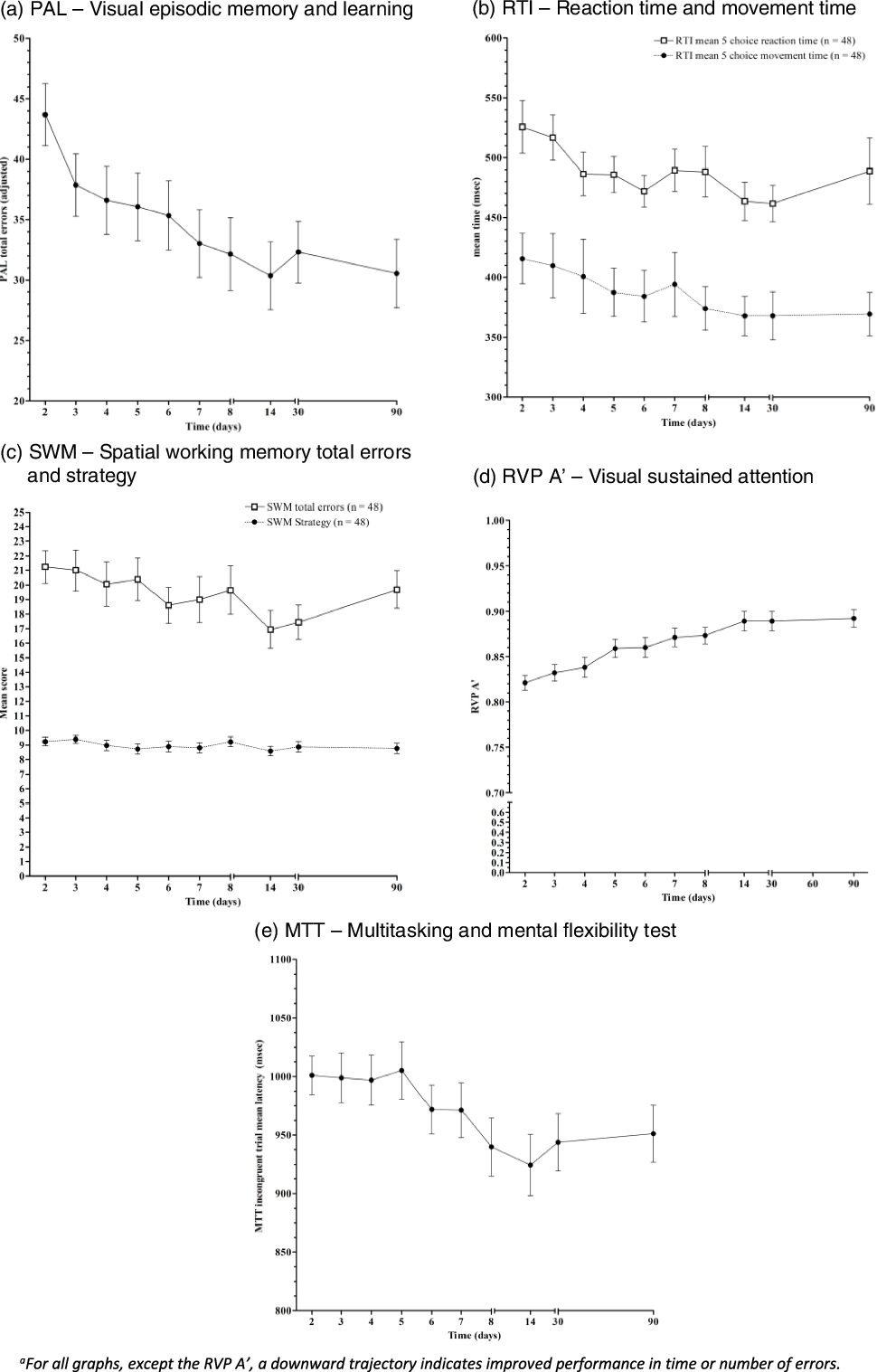
The trajectories of estimated marginal mean scores for CANTAB key measures plotted on graphs over ten timepoints from 2 to 90 days post-stroke, where all graphs show a significant trend towards improvement that stabilises from 6-14 days post-stroke, apart from the Spatial Working Memory Strategy subtest which does not show significant change.a
Time Points of Significant Change in CANTAB Key Measures*
| Time point (Days post-stroke) | 2 | 3 | 4 | 5 | 6 | 7 | 8 | 14 | 30 | 90 | |
|---|---|---|---|---|---|---|---|---|---|---|---|
| 2 | MTTLNOM | MTTLNOM | MTTLNOM | MTTLNOM | |||||||
| PALTEA | PALTEA | PALTEA | PALTEA | PALTEA | PALTEA | PALTEA | PALTEA | PALTEA | |||
| RTIFMMT | RTIFMMT | RTIFMMT | |||||||||
| RTIFMRT | RTIFMRT | RTIFMRT | RTIFMRT | RTIFMRT | RTIFMRT | ||||||
| SWMTE | SWMTE | ||||||||||
| RVPA′ | RVPA′ | RVPA′ | RVPA′ | RVPA′ | RVPA′ | RVPA′ | RVPA′ | ||||
| 3 | MTTLNOM | MTTLNOM | MTTLNOM | MTTLNOM | |||||||
| PALTEA | PALTEA | PALTEA | PALTEA | PALTEA | |||||||
| RTIFMMT | RTIFMMT | RTIFMMT | |||||||||
| RTIFMRT | RTIFMRT | RTIFMRT | RTIFMRT | ||||||||
| SWMTE | SWMTE | SWMTE | |||||||||
| RVPA′ | RVPA′ | RVPA′ | RVPA′ | RVPA′ | RVPA′ | RVPA′ | RVPA′ | ||||
| 4 | MTTLNOM | MTTLNOM | MTTLNOM | MTTLNOM | |||||||
| PALTEA | PALTEA | ||||||||||
| SWMTE | |||||||||||
| RVPA′ | RVPA′ | RVPA′ | RVPA′ | RVPA′ | RVPA′ | RVPA′ | |||||
| 5 | MTTLNOM | MTTLNOM | MTTLNOM | MTTLNOM | MTTLNOM | MTTLNOM | |||||
| PALTEA | |||||||||||
| SWMTE | SWMTE | ||||||||||
| RVPA′ | RVPA′ | RVPA′ | RVPA′ | RVPA′ | |||||||
| 6 | MTTLNOM | MTTLNOM | |||||||||
| PALTEA | |||||||||||
| -RTIFMRT | |||||||||||
| RVPA′ | RVPA′ | RVPA′ | |||||||||
| 7 | MTTLNOM | MTTLNOM | |||||||||
| RTIFMRT | RTIFMRT | ||||||||||
| RVPA′ | RVPA′ | RVPA′ | |||||||||
| 8 | |||||||||||
| SWMTE | |||||||||||
| RVPA′ | RVPA′ | RVPA′ | |||||||||
| 14 | |||||||||||
| - SWMTE | |||||||||||
| 30 | |||||||||||
| 90 |
*p < 0.05
Time points of significant change identified in post-hoc Least Square Differences for CANTAB key measures (p < .05) with: 22 significant points of change for Multitasking Test Incongruent Trial Mean Latency (MTTLNOM), a measure of mental flexibility and executive function; 18 significant points of change for Paired Associates Learning Total Errors Adjusted (PALTEA), a measure of learning and memory; 5 significant points of change for RTI Mean 5-Choice Movement Time (RTIFMMT), a measure of upper limb motor speed; 13 significant points of change for RTI Mean 5-Choice Reaction Time (RTIFMRT), a measure of reaction time; 10 significant points of change for Spatial Working Memory Total Errors (SWMTE), a measure of spatial working memory; 0 significant points of change for Spatial Working Memory Strategy (SWMS), a measure of strategy use and executive function; 37 significant points of change for Rapid Visual Processing ‘A’ Prime (RVPA’), a measure of sustained visual attention.
Paired associates learning
Repeated measures ANOVA was conducted for total errors adjusted on the Paired Associates Learning task (PALTEA) (Fig. 2) identifying a significant change over time (F(9,39) = 4.656, p. < 0.001, η2p = 0.518, power = 0.994). Post-hoc Least Square Differences comparison of timepoints identified 18 differences (p < 0.05) with performances improving significantly from day 2 to 3 with ongoing improvements until day 14 post-stroke, after which performances stabilised (Table 4). For z-score analysis, one-way repeated measures ANOVA was significant, F(9,423) = 6.104, p. < 0.001, η2p = 0.115, power = 1.0). With results otherwise identical to raw score analysis.
Five-choice reaction time
Across both five-choice RTI measures (movement time and reaction time), repeated measures MANOVA returned a significant effect of time (F(18,30) = 2.656, p. = 0.009, η2p = 0.614, power = 0.964). Follow-up repeated measures ANOVAs for each of the subtests, corrected for family-wise error, were performed. For RTI five-choice movement time (RTIFMMT), a significant change over time was found (F(9,423) = 1.883, p. = 0.053, η2p = 0.039, power = 0.828) (Fig. 2). For RTI five-choice mean reaction time (RTIFMRT), a significant change over time was also identified (F(9,423) = 3.189, p. = 0.001, η2p = 0.064, power = 0.980) (Fig. 2). Post-hoc Least Square Differences comparison of timepoints identified 13 differences (p < .05) in RTIFMRT and 5 in RTIFMMT. Movement time (RTIFMMT) performances improved steadily until day 8 post-stroke. Reaction time (RTIFMRT) showed significant improvements to day 6 after which performances stabilised (Table 4).
Spatial working memory
For SWM, a slightly different picture emerged. Repeated measures MANOVA of both SWM strategy (SWMS) and SWM total errors (SWMTE), to control for family-wise error, identified a significant effect of time (F(18,30) = 2.989, p. = 0.004, η2p = 0.642, power = 0.981). Follow-up repeated measures ANOVAs for each of the subtests corrected for family-wise error returned mixed results. For SWMS, no significant change over time was found (F(9,423) = 1.443, p. = 0.167, η2p = 0.030, power = 0.693), while for SWMTE a significant change over time was found (F(9,423) = 2.452, p. = 0.010, η2p = 0.050, power = 0.928) (Fig. 2). Post-hoc Least Square Differences comparison of timepoints identified 10 differences in SWMTE (p < 0.05) with performances improving until day 14 post-stroke, after which performances stabilised (Table 4).
Rapid visual processing
Lastly, for RVP A’, a repeated measures ANOVA identified a significant change over time (F(9,423) = 12.922, p. < 0.001, η2p = 0.749, power = 1.00) (Fig. 2). Post-hoc Least Square Differences comparison of timepoints identified 37 differences (p < 0.05) with performances improving until day 6 where they slowed slightly but continued to improve until day 14 post-stroke, after which performances stabilised (Table 4). For z-score analysis, one-way repeated measures ANOVA was significant (F(9,423) = 26.015, p. < 0.001, η2p = 0.356, power = 1.0). With results otherwise identical to raw score analysis.
In summary, post-hoc analyses of change over time for MTTLNOM, PALTEA, RTIFMRT, RTIFMMT, and RVP A’ showed a significant change within the first week of measurement, to 8 days post-stroke (Table 3). The SWMTE post-hoc analysis did not identify significant change until day 14 post-stroke (Table 3). The SWMS measure did not show any significant change (Table 3). All measures had stabilised by day 14 post-stroke. Raw mean scores are available in Table 4. Standardised scores and percentile scores are available for PALTEA and RVP A’and reveal participant’s mean scores fall below a healthy adult normative population
Discussion
Mapping the trajectory of cognitive recovery post-stroke
We found that cognitive recovery occurs steadily over the first 2 weeks post-stroke, a period we identify as post-stroke delirium, which has significant clinical implications for cognitive assessment in the acute stroke unit. Quality statements for acute stroke clinical care standards recommend that stroke survivors be assessed for rehabilitation needs, including cognitive status, within the first few days of admission to an acute stroke unit (Lindsay et al., 2014). Current practice in the acute stroke unit is screening cognition at a single time-point during a period of early, predictable global improvement (Quinn et al., 2018). Cognitive assessment results in this early phase, therefore, should be used as a marker for monitoring improvement and to direct acute clinical decisions, rather than an indicator of long-term impairment.
Monitoring cognitive improvement with pen and paper cognitive screening assessments poses a problem as alternate forms are largely not validated for serial testing over short time intervals or have few alternate versions (Costa et al., 2012; Nasreddine & Patel, 2016). In contrast, many computerised cognitive assessments, such as the CANTAB, are designed and validated for serial testing over short periods of time (Zygouris & Tsolaki, 2015); and, when used in conjunction with functional clinical assessment (Aslam et al., 2018), can be used to measure improvements during this period of acute recovery.
Our second finding is that early cognitive recovery largely stabilises by 6 to 14 days post mild stroke. Measures of visual episodic learning (PAL), impulse control and decision making (MTT), information processing speed (RTIFMRT), motor speed (RTIFMMT) and complex sustained attention (RVP) demonstrated steady significant change in the first week. Visual working memory (SWM) was slower to recover in the first week but showed significant change by day 14 when all measures stabilised. These results are consistent with a study that demonstrated generalised rather than domain specific cognitive improvement measured by traditional neuropsychological testing in the acute phase and followed up 12-24 months later (van Zandvoort et al., 2005). Additionally, differences in pace of recovery for cognitive processing speed (RTIFMRT), which significantly increased to day 6, and motor speed (RTIFMMT), which demonstrated slightly slower recovery to day 8, confirms the importance of partitioning the two during early testing. Our results indicate that early cognitive recovery begins to stabilise from days 6 to 14 in mild ischaemic stroke survivors, suggesting that formal neuropsychological assessment may occur significantly earlier than previously considered to be clinically appropriate (De Haan et al., 2006).
Domain-specific testing
Improved understanding of domain-specific patterns of recovery is clinically useful. Targeted cognitive assessment of a single domain (processing speed), found to be linked to long-term cognitive and functional impairment, has previously been suggested as time-efficient (Cumming et al., 2012). Our results indicate that specific CANTAB subtests display high levels of sensitivity to early cognitive change following mild ischaemic stroke. The RVP A’ measures complex sustained attention using a target sensitivity paradigm. Performances on RVP A’ displayed steady, consistent improvement, showing significant daily change from very early post-stroke, day 4 through day 14, with flattening of the trajectory out to 30 and 90 days. The measure displayed narrow variance across all time-points indicating a high degree of reliability of measurement.
The only measure to demonstrate no change over time was the Spatial Working Memory Strategy (SWMS) task, which involves use of strategy to minimise working memory errors when attempting to recall and manipulate visuospatial information (Cambridge Cognition, 2022). A small study of 25 participants with amnestic mild cognitive impairment found a practice effect for the SWMS task when testing was separated by 6 months (Cacciamani et al., 2018). However, our participants failed to demonstrate any significant change in score despite daily testing. Executive dysfunction is poorly predicted by stroke and is often present prior to stroke. (Heshmatollah et al., 2021; Kliper et al., 2014; Veldsman et al., 2020). Possibly this measure reflects a high degree of stable pre-stroke executive dysfunction that did not change due to the chronic nature of the impairment. The non-significance observed on this measure suggests that SWMS is a less reliable method to detect early change following mild stroke.
Post-stroke delirium
We propose that this early improvement across a broad sample of cognitive domains is evidence of a distinct state of acute stroke-induced delirium that precedes a slower neuroplastic recovery process, consistent with clinical observations of this population. This acute recovery is expected to be due to secondary effects of the stroke resolving, rather than resolution of primary effects of neuronal death, which are not likely to demonstrate such quick restitution (Gottesman & Hillis, 2010; McDonald et al., 2019).
In keeping with a delirium state, the effect of post-stroke delirium appears to be generalised across cognitive domains rather than domain specific. All CANTAB measures (Fig. 2) demonstrated pronounced cognitive improvement, with the notable exception of SWMS which did not show significant change. Other studies have revealed that when detailed neuropsychological testing is performed early post-stroke, the cognitive profile remains similar 6-24 months later, with the same constellation of impaired versus unimpaired domains (Nys et al., 2005; Wolf & Rognstad, 2013; van Zandvoort et al., 2005). Two of these studies assessed cognition 4-20 days post-stroke, and found generalised improvements, and the third study by Wolf & Rognstad (2013) assessed cognition at 3 weeks and found a stable cognitive profile. This generalised global recovery was captured when testing occurred at <3 weeks post-stroke, which is consistent with our findings. However, our findings suggest an earlier stabilisation of post-stroke delirium between 6 and 14 days post mild stroke.
Other studies have suggested that acute stroke causes a delirium-state (Maldonado, 2018; McManus et al., 2009; Stokholm et al., 2019), but acknowledged difficulty in distinguishing which factors were associated with delirium. Stroke severity is a known predictor of delirium (Oldenbeuving et al., 2011; Qu et al., 2018a), which supports the premise that stroke induces a delirium state in the acute phase. A systematic review of delirium assessment tools used in stroke and variations in reported delirium, suggested that acute stroke may cause delirium-like symptoms (Stokholm et al., 2019). A comparison of two delirium screening tools concluded that delirium detected within four days of stroke was precipitated by the stroke as no other cause was evident (Caeiro et al., 2004). Another found higher incidence of delirium in an acute stroke population when compared to acute coronary patients and concluded that delirium was likely caused by stroke (Mc Manus et al., 2009). However, these studies did not directly demonstrate early, global cognitive recovery separate from clinically recognised delirium.
One quarter of our participants had observable symptoms of delirium, captured on the 4-AT, which is validated for use in acute stroke (Lees et al., 2013; Qu et al., 2018b). This proportion is consistent with prior research reporting delirium to be present in 13-48% of stroke survivors (Oldenbeuving et al., 2007). However, using the 4-AT, most participants with symptomatic delirium appeared to resolve by day four post-stroke. When testing with a highly sensitive, computerised cognitive battery, the steady recovery we were able to measure implies ongoing resolution of this delirium state, beyond the days captured by the 4-AT. Sub-syndromal delirium has been described in the acute stroke population (Pasinska et al., 2018), which suggests that post-stroke delirium is on a continuum of severity made apparent by the more sensitive computerised testing used in our study. For mild post-stroke delirium, fluctuations in attention, a feature used in the clinical diagnosis of delirium (American Psychiatric & American Psychiatric Association, 2013), may be less apparent and therefore not captured by the delirium screening. These results illustrate that cognitive impairment is pervasive acutely post-stroke.
Strengths
Serial computerised cognitive assessment was sensitive enough to capture detailed information on domain-specific cognitive impairment and demonstrate significant recovery in a predominantly mild, ischaemic stroke cohort. These computerised assessments are highly reproducible and protect against practice effect ensuring the findings are likely to be reliable. We were also able to include stroke patients with aphasia who are often excluded in cognitive research (Wall et al., 2015). Further, we compared sensitive cognitive testing with a validated delirium screen to highlight limitations of delirium screening in a mild stroke cohort. Lastly, we gathered data up to 90 days to ensure we captured the period of greatest change post-stroke.
Limitations
Participants were classified as predominantly mild stroke and results less generalisable to a severe stroke cohort. Our selected subtests do not include assessment of language and Verbal Paired Associates could be added to address this commonly impaired domain. As well, this is a single-centre study in a dedicated acute stroke unit and results may differ in alternate settings. Lastly, our cohort of 48 participants was small and additional research with larger numbers will be required to verify these results.
Suggestions for further research
Further research is recommended to confirm the stability of domain-specific patterns of post-stroke cognitive impairment which would assist early, targeted cognitive interventions. Large studies of computerised testing could define more sensitive thresholds for identifying delirium than current screening tools. Further, exploring a broader range of computerised measures may reveal tests optimal for both screening and identification of specific clinically relevant deficits.
Some studies have found that cognition continues to recover months to years post-stroke (Hofgren et al., 2007; Hurford et al., 2012), while others demonstrate a decline in cognitive function (Jokinen et al., 2015; Levine et al., 2015). Hochstenbach et al. (2003) found that most stroke survivors showed no change in cognitive performance over a 2-year period, while some declined and others improved. The stroke survivors that improved accounted for the general upward trend of cognitive function. Further research may reveal different patterns of acute post-stroke recovery, which may be useful in predicting individual cognitive trajectories.
Conclusion
Serial computerised cognitive assessment is able to illustrate early generalised recovery over 14 days that is evidence for a distinct post-stroke delirium state. Post-stroke delirium appears to be far more common than is currently appreciated, and can be measured, even in mild stroke. Computerised cognitive assessment may be a more sensitive tool to identify delirium than current clinical screening tools. We have identified stabilisation of this acute cognitive recovery between 6 and 14 days for mild-stroke, indicating that more detailed neuropsychological testing may be performed earlier than previously understood. Further exploration of serial cognitive performance in delirium states could help us better understand this common and costly complication.
Data availability statement
The data that support the findings of this study are available on request from the corresponding author. The data are not publicly available due to privacy or ethical restrictions.
Acknowledgements
The authors gratefully thank all the participants. They acknowledge the valued contributions of the Allied Health Assistants, Christopher Duffy and Michael Lee, who assisted with data collection. The authors also thank staff of the Acute Stroke and Rehabilitation Units of the Sunshine Coast University Hospital for their support.
Authors’ contributions
AC, MS, LG and RG developed the concept and design. AC, LG, IR and RG prepared the protocol and ethics application. RG, LG and IR obtained funding. AC collected the data. MS and AC performed the statistical analysis. AC drafted the manuscript. All authors participated in the critical revisions of the manuscript.
Funding
This work was supported by the Sunshine Coast Hospital and Health Service – Transformation and Transition Funding.
Ethics approval
The authors assert that all procedures contributing to this work comply with the ethical standards of the relevant national and institutional committees on human experimentation and with the Helsinki Declaration of 1975, as revised in 2008. Ethical approval was obtained from The Prince Charles Hospital Human Research Ethics Committee, HREC/17/QPCH/163; the University of Queensland (#2017001149) and from University of the Sunshine Coast (S/17/1091).
Consent to participate
Informed consent was obtained from individual participants and/or substitute decision-makers for all participants included in the study.
Consent for publication
Participants or their substitute decision-makers signed informed consent regarding publishing their data.
References
Abzhandadze, T., Rafsten, L., Lundgren Nilsson, A., Palstam, A., Sunnerhagen, K. S. (2019) Very early MoCA can predict functional dependence at 3 months after stroke: A longitudinal, cohort study. Frontiers in Neurology [Open Access] 1-9.
| Crossref | Google Scholar |
Adamit, T., Maeir, A., Ben Assayag, E., Bornstein, N. M., Korczyn, A. D., Katz, N. (2014) Impact of first-ever mild stroke on participation at 3 and 6 month post-event: the TABASCO study. Disability and Rehabilitation 37(8), 667-673.
| Crossref | Google Scholar |
Adams, H. P., Davis, P. H., Leira, E. C., Chang, K.-C., Bendixen, B. H., Clarke, W. R., et al. (1999) Baseline NIH Stroke Scale score strongly predicts outcome after stroke. A report of the Trial of Org 10172 in Acute Stroke Treatment (TOAST) 53(1), 126.
| Crossref | Google Scholar |
Aslam, R. H. W., Bates, V., Dundar, Y., Hounsome, J., Richardson, M., Krishan, A., et al. (2018) A systematic review of the diagnostic accuracy of automated tests for cognitive impairment. International Journal of Geriatric Psychiatry 33(4), 561-575.
| Crossref | Google Scholar |
Barker-Collo, S., Feigin, V. L., Parag, V., Lawes, C. M., Senior, H. (2010) Auckland Stroke Outcomes Study. Part 2: Cognition and functional outcomes 5 years poststroke. Neurology 75(18), 1608-1616.
| Crossref | Google Scholar |
Bernhardt, J., Hayward, K. S., Kwakkel, G., Ward, N. S., Wolf, S. L., Borschmann, K., et al. (2017) Agreed definitions and a shared vision for new standards in stroke recovery research: The stroke recovery and rehabilitation roundtable taskforce. International Journal of Stroke 12(5), 444-450.
| Crossref | Google Scholar |
Bertolin, M., Van Patten, R., Greif, T., Fucetola, R. (2018) Predicting cognitive functioning, activities of daily living, and participation 6 months after mild to moderate stroke. Archives of Clinical Neuropsychology 33(5), 562-576.
| Crossref | Google Scholar |
Boulanger, J. M., Lindsay, M. P., Gubitz, G., Smith, E. E., Stotts, G., Foley, N., et al. (2018) Canadian Stroke Best Practice Recommendations for Acute Stroke Management: Prehospital, Emergency Department, and Acute Inpatient Stroke Care, 6th Edition, Update 2018. International Journal of Stroke 13(9), 949-984.
| Crossref | Google Scholar |
Brott, T., Adams, H. P., Olinger, C. P., Marler, J. R., Barsan, W. G., Biller, J., et al. (1989) Measurements of acute cerebral infarction: a clinical examination scale. Stroke 20(7), 864-870.
| Crossref | Google Scholar |
Cacciamani, F., Salvadori, N., Eusebi, P., Lisetti, V., Luchetti, E., Calabresi, P., Parnetti, L. (2018) Evidence of practice effect in CANTAB spatial working memory test in a cohort of patients with mild cognitive impairment. Applied Neuropsychology: Adult 25(3), 237-248.
| Crossref | Google Scholar |
Caeiro, L., Ferro, J. M., Albuquerque, R., Figueira, M. L. (2004) Delirium in the first days of acute stroke. Journal of Neurology 251(2), 171-178.
| Crossref | Google Scholar |
Cambridge Cognition (2021, CANTAB®, Cambridge Neuropsychological Test Automated Battery, All rights reserved. Retrieved 28 January from. Retrieved from www.cantab.com, Accessed 28 January 2021,
Cambridge Cognition (2022). CANTAB® [Cognitive assessment software], Cambridge Cognition, Retrieved 28 January from. Retrieved from www.cantab.com,
Campos-Magdaleno, M., Leiva, D., Pereiro, A. X., Lojo-Seoane, C., Mallo, S. C., Facal, D., Juncos-Rabadán, O. (2021) Changes in visual memory in mild cognitive impairment: A longitudinal study with CANTAB. Psychological Medicine 51(14), 2465-2475.
| Crossref | Google Scholar |
Chan, E., Altendorff, S., Healy, C., Werring, D. J., Cipolotti, L. (2017) The test accuracy of the Montreal Cognitive Assessment (MoCA) by stroke lateralisation. Journal of the Neurological Sciences 373(12), 100-104.
| Crossref | Google Scholar |
Chan, E., Khan, S., Oliver, R., Gill, S. K., Werring, D. J., Cipolotti, L. (2014) Underestimation of cognitive impairments by the Montreal Cognitive Assessment (MoCA) in an acute stroke unit population. Journal of the Neurological Sciences 343(1-2), 176-179.
| Crossref | Google Scholar |
Chiti, G., Pantoni, L. (2014) Use of Montreal Cognitive Assessment in patients with stroke. Stroke 45(10), 3135-3140.
| Crossref | Google Scholar |
Costa, A. S., Fimm, B., Friesen, P., Soundjock, H., Rottschy, C., Gross, T., et al. (2012) Alternate-form reliability of the Montreal Cognitive Assessment screening test in a clinical setting. Dementia and Geriatric Cognitive Disorders 33(6), 379-384.
| Crossref | Google Scholar |
Crivelli, D., Spinosa, C., Angelillo, M. T., Balconi, M. (2021) The influence of language comprehension proficiency on assessment of global cognitive impairment following acquired brain injury: A comparison between MMSE, MoCA and CASP batteries. Applied Neuropsychology: Adult 1(1), 1-6.
| Crossref | Google Scholar |
Cumming, T. B., Brodtmann, A., Darby, D., Bernhardt, J. (2012) Cutting a long story short: Reaction times in acute stroke are associated with longer term cognitive outcomes. Journal of The Neurological Sciences 322(1-2), 102-106.
| Crossref | Google Scholar |
Cumming, T. B., Marshall, R. S., Lazar, R. M. (2013) Stroke, cognitive deficits, and rehabilitation: Still an incomplete picture. International Journal of Stroke 8(1), 38-45.
| Crossref | Google Scholar |
De Haan, E., Nys, G. M., Van Zandvoort, M. J. (2006) Cognitive function following stroke and vascular cognitive impairment. Current Opinion in Neurology 19(6), 559-564.
| Google Scholar |
Duncan, P. W., Min Lai, S., Keighley, J. (2000) Defining post-stroke recovery: implications for design and interpretation of drug trials. Neuropharmacology 39(5), 835-841.
| Crossref | Google Scholar |
Gottesman, R. F., Hillis, A. E. (2010) Predictors and assessment of cognitive dysfunction resulting from ischaemic stroke. The Lancet Neurology 9(9), 895-905 https://doi.org/10.1016/S1474-4422(10)70164-2.
| Google Scholar |
Hachinski, V., Iadecola, C., Petersen, R. C., Breteler, M. M., Nyenhuis, D. L., Black, S. E., et al. (2006) National Institute of Neurological Disorders and Stroke-Canadian Stroke Network Vascular Cognitive Impairment Harmonization Standards. Stroke 37(9), 2220-2241.
| Crossref | Google Scholar |
Heshmatollah, A., Dommershuijsen, L. J., Fani, L., Koudstaal, P. J., Ikram, M. A., Ikram, M. K. (2021) Long-term trajectories of decline in cognition and daily functioning before and after stroke. Journal of Neurology, Neurosurgery & Psychiatry 92(11), 1158-1163.
| Crossref | Google Scholar |
Hochstenbach, J. B., Den Otter, R., Mulder, T. W. (2003) Cognitive recovery after stroke: A 2-year follow-up. Archives of Physical Medicine and Rehabilitation 84(10), 1499-1504.
| Crossref | Google Scholar |
Hofgren, C., Björkdahl, A., Esbjörnsson, E., Stibrant-Sunnerhagen, K. (2007) Recovery after stroke: cognition, ADL function and return to work. Acta Neurologica Scandinavica 115(2), 73-80.
| Crossref | Google Scholar |
Hurford, R., Charidimou, A., Fox, Z., Cipolotti, L., Werring, D. J. (2012) Domain-specific trends in cognitive impairment after acute ischaemic stroke. Journal of Neurology 260(1), 237-241.
| Crossref | Google Scholar |
Jokinen, H., Melkas, S., Ylikoski, R., Pohjasvaara, T., Kaste, M., Erkinjuntti, T., Hietanen, M. (2015) Post-stroke cognitive impairment is common even after successful clinical recovery. European Journal of Neurology 22(9), 1288-1294.
| Crossref | Google Scholar |
Kliper, E., Ben Assayag, E., Tarrasch, R., Artzi, M., Korczyn, A. D., Shenhar-Tsarfaty, S., et al. (2014) Cognitive state following stroke: The predominant role of preexisting white matter lesions. PLoS ONE 9(8), e105461.
| Crossref | Google Scholar |
Koski, L. (2013) Validity and applications of the Montreal Cognitive Assessment for the assessment of vascular cognitive impairment. Cerebrovascular Diseases 36(1), 6-18.
| Crossref | Google Scholar |
Krishnamurthi, R., Ikeda, T., Feigin, V. (2020) Global, regional and country-specific burden of ischaemic stroke, intracerebral haemorrhage and subarachnoid haemorrhage: A systematic analysis of the global burden of disease study 2017. Neuroepidemiology 54(Suppl. 2), 171-179.
| Crossref | Google Scholar |
Lanctôt, K. L., Lindsay, M. P., Smith, E. E., Sahlas, D. J., Foley, N., Gubitz, G., et al. (2020) Canadian stroke best practice recommendations: Mood, cognition and fatigue following stroke, update 2019. International Journal of Stroke 15(6), 668-688.
| Crossref | Google Scholar |
Lees, R., Corbet, S., Johnston, C., Moffitt, E., Shaw, G., Quinn, T. J. (2013) Test accuracy of short screening tests for diagnosis of delirium or cognitive impairment in an acute stroke unit setting. Stroke 44(11), 3078-3083.
| Crossref | Google Scholar |
Levine, D. A., Galecki, A. T., Langa, K. M., Unverzagt, F. W., Kabeto, M. U., Giordani, B., Wadley, V. G. (2015) Trajectory of cognitive decline after incident stroke. JAMA 314(1), 41-51.
| Crossref | Google Scholar |
Lindsay, P., Furie, K. L., Davis, S., Donnan, G., Norrving, B. (2014) World stroke organization global stroke services guidelines and action plan. International Journal of Stroke 9(SA100), 4-13.
| Crossref | Google Scholar |
Lo, J. W., Crawford, J. D., Desmond, D. W., Bae, H.-J., Lim, J.-S., Godefroy, O., et al. (2021) Long-term cognitive decline after stroke: An individual participant data meta-analysis. Stroke 53(4), 1318-1327.
| Crossref | Google Scholar |
Mahon, S., Parmar, P., Barker-Collo, S., Krishnamurthi, R., Jones, K., Theadom, A., Feigin, V. (2017) Determinants, prevalence, and trajectory of long-term post-stroke cognitive impairment: Results from a 4-year follow-up of the ARCOS-IV study. Neuroepidemiology 49(3-4), 129-134.
| Crossref | Google Scholar |
Maldonado, J. R. (2018) Delirium pathophysiology: An updated hypothesis of the etiology of acute brain failure. International Journal of Geriatric Psychiatry 33(11), 1428-1457.
| Crossref | Google Scholar |
Mc Manus, J., Pathansali, R., Hassan, H., Ouldred, E., Cooper, D., Stewart, R., et al. (2009) The evaluation of delirium post-stroke. International Journal of Geriatric Psychiatry 24(11), 1251-1256.
| Crossref | Google Scholar |
McDonald, M. W., Black, S. E., Copland, D. A., Corbett, D., Dijkhuizen, R. M., Farr, T. D., et al. (2019) Cognition in stroke rehabilitation and recovery research: Consensus-based core recommendations from the second Stroke Recovery and Rehabilitation Roundtable. International Journal of Stroke 14(8), 774-782.
| Crossref | Google Scholar |
McManus, J., Pathansali, R., Hassan, H., Ouldred, E., Cooper, D., Stewart, R., et al. (2009) The evaluation of delirium post-stroke. International Journal of Geriatric Psychiatry 24(11), 1251-1256.
| Crossref | Google Scholar |
Mole, J. A., Demeyere, N. (2020) The relationship between early post-stroke cognition and longer term activities and participation: A systematic review. Neuropsychological Rehabilitation 30(2), 346-370.
| Crossref | Google Scholar |
Nasreddine, Z. S., Patel, B. B. (2016) Validation of Montreal Cognitive Assessment, MoCA, alternate french versions. Canadian Journal of Neurological Sciences / Journal Canadien des Sciences Neurologiques 43(5), 665-671.
| Crossref | Google Scholar |
Nasreddine, Z. S., Phillips, N. A., Bã©Dirian, V. R., Charbonneau, S., Whitehead, V., Collin, I., et al. (2005) The montreal cognitive assessment, MoCA: A brief screening tool for mild cognitive impairment. Journal of the American Geriatrics Society 53(4), 695-699.
| Crossref | Google Scholar |
Ng, F., Trauer, T., Dodd, S., Callaly, T., Campbell, S., Berk, M. (2007) The validity of the 21-item version of the Depression Anxiety Stress Scales as a routine clinical outcome measure. Acta Neuropsychiatrica 19(5), 304-310.
| Crossref | Google Scholar |
Nys, G. M. S., Van Zandvoort, M. J. E., De Kort, P. L. M., Jansen, B. P. W., Van Der Worp, H. B., Kappelle, L. J., De Haan, E. H. F. (2005) Domain-specific cognitive recovery after first-ever stroke: A follow-up study of 111 cases. Journal of the International Neuropsychological Society 11(07), 339.
| Crossref | Google Scholar |
Nys, G. M. S., Van Zandvoort, M. J. E., Van Der Worp, H. B., De Haan, E. H. F., De Kort, P. L. M., Jansen, B. P. W., Kappelle, L. J. (2006) Early cognitive impairment predicts long-term depressive symptoms and quality of life after stroke. Journal of the Neurological Sciences 247(2), 149-156.
| Crossref | Google Scholar |
Oldenbeuving, A. W., de Kort, P. L., Jansen, B. P., Algra, A., Kappelle, L. J., Roks, G. (2011) Delirium in the acute phase after stroke: incidence, risk factors, and outcome. Neurology 76(11), 993-999.
| Crossref | Google Scholar |
Oldenbeuving, A. W., De Kort, P. L. M., Jansen, B. P. W., Roks, G., Kappelle, L. J. (2007) Delirium in acute stroke: A review. International Journal of Stroke 2(4), 270-275.
| Crossref | Google Scholar |
Pasinska, P., Kowalska, K., Klimiec, E., Szyper-Maciejowska, A., Wilk, A., Klimkowicz-Mrowiec, A. (2018) Frequency and predictors of post-stroke delirium in PRospective Observational POLIsh Study (PROPOLIS). Journal of Neurology 265(4), 863-870.
| Crossref | Google Scholar |
Pendlebury, S. T., Klaus, S. P., Thomson, R. J., Mehta, Z., Wharton, R. M., Rothwell, P. M., et al. (2015) Methodological factors in determining risk of dementia after transient ischemic attack and stroke: (III) Applicability of cognitive tests. Stroke 46(11), 3067-3073.
| Crossref | Google Scholar |
Pettigrew, C., Soldan, A., Brichko, R., Zhu, Y., Wang, M.-C., Kutten, K., et al. (2021) Computerized paired associate learning performance and imaging biomarkers in older adults without dementia. Brain Imaging and Behavior 16(2), 921-929.
| Crossref | Google Scholar |
Qu, J., Chen, Y., Luo, G., Zhong, H., Xiao, W., Yin, H. (2018) Delirium in the acute phase of ischemic stroke: Incidence, risk factors, and effects on functional outcome. Journal of Stroke and Cerebrovascular Diseases 27(10), 2641-2647.
| Crossref | Google Scholar |
Qu, J., Chen, Y., Luo, G., Zhong, H., Xiao, W., Yin, H. (2018) Delirium in the acute phase of ischemic stroke: Incidence, risk factors, and effects on functional outcome. Journal of Stroke and Cerebrovascular Diseases 27(10), 2641-2647.
| Crossref | Google Scholar |
Quinn, T. J., Elliott, E., Langhorne, P. (2018) Cognitive and mood assessment tools for use in stroke. Stroke 49(2), 483-490.
| Crossref | Google Scholar |
Quinn, T. J., Richard, E., Teuschl, Y., Gattringer, T., Hafdi, M., O’Brien, J. T., et al. (2021) European stroke organisation and European Academy of Neurology joint guidelines on post-stroke cognitive impairment. European Journal of Neurology 28(12), 3883-3920.
| Crossref | Google Scholar |
Shopin, L., Shenhar-Tsarfaty, S., Ben Assayag, E., Hallevi, H., Korczyn, A. D., Bornstein, N. M., Auriel, E. (2013) Cognitive assessment in proximity to acute ischemic stroke/transient ischemic attack: comparison of the Montreal Cognitive Assessment Test and MindStreams computerized cognitive assessment battery. Dementia and Geriatric Cognitive Disorders 36(1-2), 36-42.
| Crossref | Google Scholar |
Skirrow, C., Cashdollar, N., Granger, K., Jennings, S., Baker, E., Barnett, J., Cormack, F. (2021) Test-retest reliability on the Cambridge Neuropsychological Test Automated Battery: Comment on Karlsen et al., (2020). Applied Neuropsychology: Adult 29(5), 889-892.
| Crossref | Google Scholar |
Steward, K. A., Novack, T. A., Kennedy, R., Crowe, M., Marson, D. C., Triebel, K. L. (2016) The Wechsler test of adult reading as a measure of premorbid intelligence following traumatic brain injury. Archives of Clinical Neuropsychology : the Official Journal of the National Academy of Neuropsychologists 72, ES1.
| Google Scholar |
Stokholm, J., Steenholt, J. V., Csilag, C., Kjær, T. W., Christensen, T. (2019) Delirium assessment in acute stroke: A systematic review and meta-analysis of incidence, assessment tools, and assessment frequencies. Journal of Central Nervous System Disease 11, 117957351989708.
| Crossref | Google Scholar |
Stolwyk, R. J., O’Neill, M. H., McKay, A. J. D., Wong, D. K. (2014) Are cognitive screening tools sensitive and specific enough for use after stroke? Stroke 45(10), 3129-3134.
| Crossref | Google Scholar |
Stroke Foundation (2022). Clinical Guidelines for Stroke Management. Retrieved March 13 from. Retrieved from https://informme.org.au/en/Guidelines/Clinical-Guidelines-for-Stroke-Management
Tatemichi, T. K., Desmond, D. W., Stern, Y., Paik, M., Sano, M., Bagiella, E. (1994) Cognitive impairment after stroke: frequency, patterns, and relationship to functional abilities. Journal of Neurology, Neurosurgery and Psychiatry 57,.
| Crossref | Google Scholar |
Tellier, M., Rochette, A. (2015) Falling through the cracks: A literature review to understand the reality of mild stroke survivors. Topics in Stroke Rehabilitation 16(6), 454-462.
| Crossref | Google Scholar |
van Zandvoort, M. J. E., Kessels, R. P. C., Nys, G. M. S., de Haan, E. H. F., Kappelle, L. J. (2005) Early neuropsychological evaluation in patients with ischaemic stroke provides valid information. Clinical Neurology and Neurosurgery 107(5), 385-392.
| Crossref | Google Scholar |
Veldsman, M., Werden, E., Egorova, N., Khlif, M. S., Brodtmann, A. (2020) Microstructural degeneration and cerebrovascular risk burden underlying executive dysfunction after stroke. Scientific Reports 10(1), 269.
| Crossref | Google Scholar |
Verdelho A., Wardlaw J., Pavlovic A., Pantoni L., Godefroy O., Duering M., Charidimou A., Chabriat H., Biessels G. J. (2021) Cognitive impairment in patients with cerebrovascular disease: A white paper from the links between stroke ESO Dementia Committee. European Stroke Journal 6(1), 5-17.
| Crossref | Google Scholar |
Wall, K. J., Isaacs, M. L., Copland, D. A., Cumming, T. B. (2015) Assessing cognition after stroke. Who misses out? A systematic review. International Journal of Stroke 10(5), 665-671.
| Crossref | Google Scholar |
Wolf, T. J., Rognstad, M. C. (2013) Changes in cognition following mild stroke. Neuropsychological Rehabilitation 23(2), 256-266.
| Crossref | Google Scholar |
Zheng, F., Yan, L., Zhong, B., Yang, Z., Xie, W. (2019) Progression of cognitive decline before and after incident stroke. Neurology 93(1), e20-e28.
| Crossref | Google Scholar |
Zietemann, V., Georgakis, M. K., Dondaine, T., Müller, C., Mendyk, A.-M., Kopczak, A., et al. (2018) Early MoCA predicts long-term cognitive and functional outcome and mortality after stroke. Neurology 91(20), e1838-e1850.
| Crossref | Google Scholar |
Zygouris, S., Tsolaki, M. (2015) Computerized cognitive testing for older adults. American Journal of Alzheimer’s Disease & Other Dementiasr 30(1), 13-28.
| Crossref | Google Scholar |