

Glucose consumption and gene expression in granulosa cells collected before and after in vitro oocyte maturation in the southern white rhinoceros (Ceratotherium simum simum)
E. Ruggeri A * , C. Young A , N. Ravida A , M. A. Sirard B , R. Krisher C , M. de la Rey D , C. Herbst D and B. Durrant A
A Reproductive Sciences, Beckman Center for Conservation Research, San Diego Zoo Wildlife Alliance, 15600 San Pasqual Valley Road, Escondido, CA 92027, USA.
B Departement des Sciences Animales, Centre de Recherce en Reproduction, Développement et Santé Inter-générationnelle (CRDSI), Université Laval, Pavillion Des Services, Local 2732, Quebec, QC G1V 0A6, Canada.
C Genus PLC, 1525 River Road, De Forest, WI 53532, USA.
D Embryo Plus, 41 Hendrik Vrewoerd Avenue, Brits 0250, South Africa.
Reproduction, Fertility and Development 34(13) 875-888 https://doi.org/10.1071/RD22071
Published online: 25 July 2022
© 2022 The Author(s) (or their employer(s)). Published by CSIRO Publishing. This is an open access article distributed under the Creative Commons Attribution-NonCommercial-NoDerivatives 4.0 International License (CC BY-NC-ND)
Abstract
Context: With two northern white rhinos (NWR) remaining, the continued existence of this species relies on studying their relative, the southern white rhino (SWR).
Aims: (1) Characterise gene expression in granulosa cells (GC) from SWR cumulus oocyte complexes (COCs) prior to (Pre-) and after (Post-) in vitro maturation (IVM), comparing culture media and oocytes from donors treated with or without gonadotropin stimulation prior to ovum recovery; and (2) evaluate COC glucose consumption in spent media.
Methods: COCs were retrieved from four SWRs. Granulosa cells were collected before and after IVM in SDZ or IZW medium. Total RNA was evaluated by qPCR.
Key results: Oocyte maturation was greater in SDZ than IZW media. Expression of genes associated with follicle development increased in Pre-IVM GC. Six genes were differentially expressed in Post-IVM GC from stimulated compared to unstimulated donors. COCs from stimulated animals consumed more glucose. Fifty seven percent of oocytes in SDZ medium consumed all available glucose.
Conclusions: Gene expression changed upon in vitro maturation and gonadotropin stimulation. Higher glucose availability might be needed during IVM.
Implications: This is the first study examining GC gene expression and COC metabolic requirements in rhinoceros, which are critical aspects to optimise IVM of rhinoceros oocytes.
Keywords: assisted reproductive technologies, endangered species, granulosa cell, in vitro maturation, oocyte, ovum pickup, reproduction, rhinoceros.
Introduction
With only two female individuals left (currently located in Kenya at Ol Pejeta Conservancy), and limited opportunities to collect their oocytes, there is little time to rescue the northern white rhino from extinction. Although some success in oocyte collection via ovum pick up (OPU), in vitro fertilisation and embryo production have been communicated (https://www.olpejetaconservancy.org/newly-created-embryo) for the last two living northern white rhinos, the methods are not yet routinely successful, and several aspects of the species’ reproductive physiology are still undetermined. Currently, there are limited opportunities to collect in vivo oocytes from any rhino species due to management challenges and the technical difficulties of performing OPU in these animals. As the closest relative to the northern white rhino, the southern subspecies is the best model for optimising the assisted reproductive technologies (ARTs) needed to save the critically endangered northern form.
While supporting oocyte competence acquisition and follicle growth, mural granulosa cells and cumulus cells play a central role in directing oocyte maturation and fertilisation potential. Analysing these specialised cells provides insight into the follicular development and differentiation process associated with oocyte quality in mammalian species. Recent transcriptome studies in the domestic cow described granulosa cell dynamics in different physiological stages to understand the physiology of ovarian and follicular changes (Khan et al. 2016). The study demonstrated that granulosa cells in a large, dominant follicle are characterised by a high level of LH receptor (LHR). During the growth phase of folliculogenesis there is a high abundance of FSH receptor (FSHR), and when a follicle becomes atretic there is a significant decrease in LHR, whereas FSHR remains unchanged. Furthermore, this work established a collection of candidate genes extensively involved in each follicular stage in the cow, giving access to a broad platform for gene expression profiles in granulosa cells.
Although there is evidence in several species (Sugiura et al. 2005; Kuo et al. 2011; Douville and Sirard 2014; Girard et al. 2015; Munakata et al. 2016) of the involvement of granulosa cells for optimal oocyte competence during folliculogenesis, limited literature has focused on determining the time and stage specific changes occurring in these cells that impact oocyte quality. In the domestic horse model, a species with physiology similar to the rhinoceros, a microarray analysis of granulosa cells was performed on dominant follicles determining distinct gene expression profiles and molecular pathways associated with developmental stages (Donadeu et al. 2014). In this study several differentially expressed transcripts were identified as unique to equine granulosa cells associated with follicular development compared to other species.
Within the bovine (Boni 2012) and equine industries (Galli et al. 2014), ovarian stimulation is a common practice prior to ovum pick up (Bols and Stout 2018). Studies have shown that ovarian stimulation has an impact on gene expression in the granulosa cells (Khan et al. 2016). Moreover, ovarian stimulation activates genes involved in oxidative stress response and growth-related genes (Dias et al. 2013), such as glutamine-fructose-6-phosphate transaminase 2 (GFPT2), which is upregulated in granulosa cells from stimulated animals. Collectively, literature from several other species reported above support our interest in analysing gene expression in granulosa cells using gene candidates involved in follicular development, oxidative stress, and metabolism.
Along with granulosa cell gene expression analysis, we sought to evaluate some aspects of oocyte metabolism during in vitro maturation. Energy metabolism is crucial for oocyte maturation and meiotic competence acquisition. It has been suggested that a substantial amount of energy is necessary for the oocyte to mature (Xie et al. 2016). Glucose is one of the main substrates used by the COC to achieve maturation (Lewis et al. 2020) and it is utilised in glycolysis as well as pentose phosphate, hexosamine biosynthesis, and polyol pathways (Sutton-McDowall et al. 2010). Importantly, gonadotropin-induced meiosis and progression to metaphase II in oocytes depends on the presence of glucose and its metabolism (Xie et al. 2016). In the mouse (Harris et al. 2009), pig (Sturmey and Leese 2003), and cow (Sutton et al. 2003b) granulosa cells perform glycolysis to generate ATP for their own use and generate pyruvate as an energy source for the oocyte. In the equine oocyte, aerobic glycolysis is the primary source of glucose metabolism during in vitro maturation (Lewis et al. 2020) and is correlated to follicular growth and oocyte maturation (Gérard et al. 2000). The oocyte is very sensitive to glucose concentration-dependent perturbations, and too much or too little glucose can lead to premature resumption of nuclear maturation (Sutton et al. 2003a).
Overall, evaluating granulosa cell gene expression and glucose metabolism will increase understanding of physiological changes in a diverse pool of follicles and the metabolic requirements of rhino COC to undergo meiotic progression and reach competence. The goal of this study was to determine both metabolic and gene expression changes in GC prior to and after in vitro maturation from donors with or without gonadotropin stimulation. We utilised granulosa cells to gain information on gene expression associated with in vitro maturation and to investigate glucose uptake during in vitro maturation. Although this is a preliminary study, this is the first research to evaluate gene expression in granulosa cells and COC glucose uptake in the southern white rhino.
Materials and methods
Animal management and ovarian stimulation protocol
This research was conducted at San Diego Zoo Wildlife Alliance’s Safari Park in Escondido, CA. Two of the four SWR oocyte donors received a single dose of 1.8 mg of Deslorelin (Sucromate, Thorn Bioscience LLC), a gonadotropin releasing hormone (GnRH) analog. The single dose was administered at an unknown stage of the estrous cycle and oocyte retrieval was performed 24 h after injection.
Anesthetic protocol and oocyte retrieval via transrectal ovum pick up (OPU)
All rhinos (n = 4) were anesthetised with a combination of etorphine (1.5–1.9 mcg/kg), butorphanol (14–39 mcg/kg), and medetomidine (20–31 mcg/kg); two of the rhinos also received azaperone (23–27 mcg/kg) and a third received midazolam (15 mcg/kg) administered intramuscularly. All four rhinos required propofol administered i.v. during initial positioning for intubation (300–658 mcg/kg) with additional supplementation required for the only rhino that did not receive any midazolam or azaperone during induction after 1 hour of anesthesia (additional 821 mcg/kg). The rhinos were reversed with atipamezole delivered i.m. at a 5:1 ratio to medetomidine (100–136 mcg/kg) and naltrexone delivered i.m. at a target 50:1 ratio to etorphine (80–110 mcg/kg) or atipamezole i.m. at a 7.5:1 ratio to medetomidine (231 mcg/kg) and naltrexone i.v. at 50:1 ratio to etorphine (77 mcg/kg).
Oocyte retrieval was executed rectally following fecal removal, rinsing and disinfection. The OPU instrument was a customised, ultrasound-guided probe with three double lumen needles. Each visible follicle was aspirated and rinsed with a flushing solution (Vigro) kept at 37°C with 12.5 I.U./mL of heparin. The follicle aspirate was kept at 37°C during transport to the laboratory where it was filtered through a 0.22 μm embryo filter (Professional Embryo Transfer Supply Inc.). The filter was rinsed with flush medium (ABT complete flush; ABT360) to isolate COCs.
Oocyte in vitro maturation (IVM)
Cumulus oocyte complexes were located using a dissecting microscope at 20×, washed with Vigro holding medium (Vetoquinol) and held at 22°C for 6–11 h. COCs were cultured in 30 μL drops singly or in pairs under light mineral oil (Global) at 38.5°C in a humidified atmosphere of 6% CO2, for 36–38 h in one of two maturation media (SDZ [M199 earls, 5 mU/mL FSH, 10.6 μg/mL LH, 1 μg/mL Estradiol, 10 ng/mL EGF, 1 mU/mL Somatotropin, 10% FBS, 9% horse follicular fluid, 50 μg/mL gentamicin] or IZW [DMEM/F12, 10% rhino estrus serum, 50 μg/mL gentamicin (Hildebrandt et al. 2018)]). After maturation, cumulus cells were denuded by pipetting in 500 U/mL hyaluronidase and ICSI was performed on all oocytes, regardless of polar body extrusion.
Sperm selection and intracytoplasmic sperm injection (ICSI) and in vitro embryo culture (IVC)
Frozen semen from a SWR bull of proven fertility was thawed in a 37°C water bath for 60 s. Sperm was layered under M199 hanks, 0.003 g/mL BSA, 50 μg/mL gentamicin and allowed to swim up for 20 min. Two microliters of sperm suspension were placed in a 5 μL drop of a commercial 10% PVP solution (Irvine Scientific) on a manipulation plate. Intracytoplasmic sperm injection (ICSI) was performed with a standard ICSI pipette. After sperm injection, oocytes were placed singly or in groups of 2 in 30 μL droplets of embryo culture medium (Global, 10% FBS) under light mineral oil and maintained at 38.5°C in a humidified atmosphere under mixed gas, 7% CO2, 6% O2, 87% N2. Cleavage was assessed on day 3 after ICSI. Embryo culture was discontinued after day 3 due to lack of embryo development.
Glucose consumption analysis
Sixteen COCs used to test glucose consumption were matured in vitro singularly in culture drops of 30 μL under oil for an average of 36 h. The total glucose available in the maturation medium was determined by evaluating the complete formulation of the culture medium.
d-glucose concentration present in maturation medium before and after COC culture was determined with a glucose colorimetric assay kit (BioVision) per the manufacturer’s instructions. Briefly, 2 μL of glucose probe, 2 μL of enzyme mix and 46 μL of assay buffer were incubated with 2 μL sample at 37°C for 30 min protected from light. After incubation, samples were read on an SpectraMax iD3 microplate reader. All samples were run in triplicate. The concentration of glucose in each sample was determined based on a standard curve prepared with the sample reactions. The glucose concentration in mg/dL was determined averaging the concentration of the triplicate wells, dividing by the volume of sample analysed and multiplying by the dilution factor. Glucose consumption during maturation was determined by subtracting the concentration of glucose in the spent media from the concentration of glucose in the media prior to culture. The glucose consumption in pmol/COC/h was determined by multiplying the amount of glucose consumed by the volume of the maturation drop (30 μL), then dividing by the length of maturation (36 h).
Granulosa cell RNA isolation and cDNA synthesis
Free-floating mural granulosa cells collected at OPU were considered in vivo cells and were identified as ‘Pre-IVM’, n = 2. These two granulosa cell samples were obtained from two different females, one of which received ovarian follicle stimulation. Granulosa cells were collected from 15 of the 22 COCs obtained by OPU. Two of these COCs were cultured together in a single drop. These granulosa cells were removed from the analysis of maturation status, as it was not possible to determine their source. Granulosa cells (cumulus) from all four females were collected after in vitro maturation from COCs cultured in single drops and were identified as Post-IVM, n = 14.
Non-atretic granulosa cells were selected prior to IVM (mural) and by manually stripping the COCs after ∼36 h of IVM culture (cumulus). Total RNA was isolated from granulosa cells using the Arcturus PicoPure RNA Isolation Kit (Thermo Fisher Scientific) per the manufacturer’s instructions. Briefly, granulosa cells were incubated with extraction buffer for 30 min before centrifugation to remove debris and extracellular material. The cell extract was incubated with ethanol and washed through pre-conditioned columns. Total RNA was eluted into extraction buffer and quantified using a Nanodrop (Thermo Scientific). Reverse transcription was performed using a QuantaBio qScript cDNA Synthesis Kit (VWR). Total RNA was added to each reverse transcription reaction with 4 μL of reaction mix, 1 μL of reverse transcriptase, and nuclease-free water to reach a total reaction volume of 20 μL. cDNA synthesis was performed at 22°C for 5 min, 42°C for 30 min, 85°C for 5 min and held at 4°C.
Quantitative real time polymerase chain reaction (qPCR) on granulosa cells
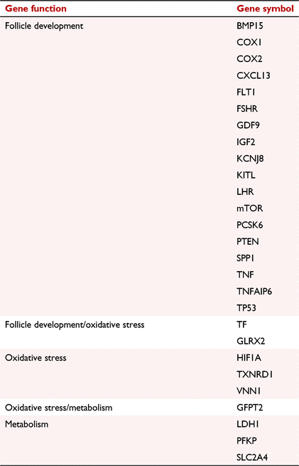
Southern white rhinoceros specific forward and reverse primers were designed using Primer3 (http://primer3.wi.mit.edu/) with a product size between 75–200 base pairs (bp), a primer length between 18–22 bp, a primer annealing temperature between 58–62°C, and a GC content between 50–60%. Primer sequences for each gene are listed in Supplemental Table 1. Genes were chosen based upon their function in cellular function, i.e. follicle development, oxidative stress, and/or metabolism. Genes and categories are listed in Table 1.
|
Twenty-seven genes were chosen for qPCR analysis based upon their functions in granulosa cell and oocyte maturation. The genes selected based upon follicle development functions were: bone morphogenetic protein 15 (BMP15), cyclooxygenase-1 (COX-1), cyclooxygenase-2 (COX-2), C-X-C motif chemokine ligand 13 (CXCL13), FMS related receptor tyrosine kinase 1 (FLT1), follicle stimulating hormone receptor (FSHR), growth differentiation factor 9 (GDF9), insulin like growth factor 2 (IGF2), potassium inwardly rectifying channel subfamily J member 8 (KCNJ8), kit ligand (KITL), luteinising hormone receptor (LHR), mechanistic target of rapamycin kinase (mTOR), proprotein convertase subtilisin/kexin type 6 (PCKS6), phosphatase and tensin homolog (PTEN), secreted phosphoprotein 1 (osteopontin/SPP1), tumor necrosis factor (TNF), tumor necrosis factor alpha induced protein 6 (TNFAIP6), and tumor protein p53 (TP53). The genes selected for oxidative stress were: glutaredoxin 2 (GLRX2), hypoxia inducible factor 1 subunit alpha (HIF1A), thioredoxin reductase 1 (TXNRD1), and vanin 1 (VNN1). The genes selected for metabolism were: lactate dehydrogenase A (LDH1), phosphofructokinase platelet (PFKP), and solute carrier family 2 member 4 (SLC2A4). A few genes overlapped in their functions. These genes included transferrin (TF) for follicle development and oxidative stress, and glutamine-fructose-6-phosphate transaminase 2 (GFPT2) for oxidative stress and metabolism.
Synthesised cDNA was utilised for quantitative real time PCR (qPCR). Bio-Rad iTaq Universal SYBR Green Supermix was used for each reaction. Each qPCR reaction contained 1.24 ng of cDNA, 2 μL of primer mix, water and 10 μL SYBR Green Supermix to reach a total volume of 20 μL. Samples were loaded into 96 well MicroAmp Fast Optical Reaction Plates (ThermoFisher Scientific) and analysed in duplicate using QuantStudio6. qPCR cycle conditions were per the manufacturer’s protocol: initial denaturation at 95°C for 5 min, 40 cycles of denaturation at 95°C for 15 s and annealing/extension at 60°C for 60 s and melt curve analysis at 65–95°C for 15 s at 0.05°C per second. Cycle threshold (CT) values were normalised to an internal control, glutathione peroxidase 7.
Statistical analyses
All statistical analyses were performed and graphed with GraphPad Prism (GraphPad Software). The data obtained from the glucose assay was analysed with an unpaired, two-tailed student’s t-test for unequal variances. For qPCR analyses, normalised CT values were utilised for statistical comparisons. Differences in Pre-IVM and Post-IVM granulosa cell gene expression were analysed with a two-way ANOVA with the Tukey test applied to correct for multiple comparisons. For comparisons between groups and animals, an unpaired two-tailed student’s t-test was utilised. The Benjamini–Hochberg method for controlling false discovery rates (FDR) was applied for multiple comparisons of 27 genes, and to decrease the false discovery rate due to the low sample number. For all statistical analyses, samples were considered statistically different at P ≤ 0.05. All data were transformed to 2−ΔCT for graphical representation. Fold changes were calculated as log2(Group A/Group B). If the fold change was positive, it indicated that gene expression was higher in group A. If the fold change was negative, it indicated that gene expression was higher in group B.
Results
Oocyte recovery and in vitro maturation rates
In the first successful OPU attempt in southern white rhinos in North America, we successfully collected 22 COCs at the San Diego Zoo Wildlife Alliance’s Safari Park (SDZWA). Two of the four females were stimulated with a GnRH agonist prior to OPU. A total of five COCs were collected from the two stimulated females, and 17 COCs were obtained from the two unstimulated females. Regardless of stimulation status, of the 22 COCs retrieved 11 were matured in SDZ medium and 11 in IZW medium. Maturation was achieved in 45% (5/11) of the oocytes cultured in SDZ medium, whereas 9% (1/11) of the oocytes cultured in IZW medium achieved maturation. One oocyte matured in SDZ medium cleaved after ICSI (1/5, 20%), and no blastocysts developed.
Effect of ovarian stimulation on granulosa cell gene expression
To determine if ovarian follicle stimulation altered the expression of the candidates’ genes chosen for analysis, differences between cells from stimulated and unstimulated females were evaluated Pre-IVM (Fig. 1). Of the 11 genes differentially expressed (COX1, FLT1, FSHR, KCNJ8, KITL, LHR, SPP1, GLRX2, VNN1, PFKP and SLC2A4I, the majority (8/11) were involved in follicle development. All gene expression averages and standard errors can be found in Supplemental Table 2.

|
Initially, gene expression of Post-IVM cells from stimulated and unstimulated animals was compared regardless of medium type. Six genes were differentially expressed; three involved in follicle development (FSHR, KCNJ8 and KITL), one in oxidative stress (VNN1) and two in metabolism (LDH1, PFKP) (Fig. 2). The effect of media formulation on gene expression in granulosa cells from stimulated and unstimulated females is demonstrated in Supplemental Fig. 1. In SDZ medium, three genes were differentially expressed between cells from stimulated and unstimulated females (FSH, GLRX2 and LDH1; Supplemental. Fig. 1a). Interestingly, there were no differentially expressed genes between stimulated and unstimulated females when cells were cultured in IZW medium, supporting the importance of the follicular status prior to OPU (Supplemental Fig. 1b).

|
Effect of in vitro maturation medium on granulosa cell gene expression
The effect of maturation medium formulation on the expression of candidate genes involved in follicle development, oxidative stress, and metabolism was examined. No significant differences were found in gene expression of granulosa cells after in vitro maturation in SDZ or IZW media (Fig. 3). Further analysis compared medium-specific gene expression changes in granulosa cells before and after in vitro maturation (Fig. 4). Two genes were differentially expressed in Pre-IVM and Post-IVM cells cultured in SDZ medium: PTEN (P = 0.03), involved in follicle development, and TXNRD1 (P = 0.03), involved in oxidative stress (Fig. 4a). Interestingly, six genes were differentially expressed before and after in vitro maturation in IZW medium (Fig. 4b). Of these genes, five were involved in follicle development (COX2, FLT1, PCSK6, TNF and TNFAIP6) and one gene in oxidative stress (HIF1A).

|

|
To investigate possible gene expression variations attributable to maturation medium formulation, mature oocytes with visible polar bodies were analysed (Fig. 5). A single gene, HIF1A (P = 0.02), was differentially expressed.

|
Oocyte glucose consumption during in vitro maturation
The glucose available was determined to be 4.54 mM for SDZ medium and 15.4 mM for IZW medium. The mean overall consumption of glucose in rhino oocytes matured in vitro, regardless of maturation medium, was 3.86 ± 2.65 mM.
Effect of ovarian stimulation on oocyte glucose consumption
To assess the effect of ovarian stimulation on the metabolism of oocytes matured in vitro, we compared glucose consumption in spent medium in COCs collected from female rhinos receiving GnRH stimulation (n = 3 COCs) and those that did not (n = 13 COCs). Due to limitations in the glucose assay from oil interference in the drops and four COCs being co-cultured, not all drops were tested for spent medium analysis. Our results indicated that ovarian stimulation caused an increase in glucose consumption during IVM regardless of culture medium used (P = 0.034; Fig. 6). Due to low numbers of COCs cultured in SDZ medium (n = 1 from stimulated females, and n = 6 from unstimulated females), statistical analysis based on ovarian stimulation was not possible. For IZW medium, COCs from stimulated animals (n = 2) consumed more glucose than those from unstimulated animals (n = 7; P = 0.007; Fig. 7).

|

|
Effect of media formulation on oocyte glucose consumption
To determine the effect of different culture media on COC glucose consumption during in vitro maturation, we compared COCs matured in SDZ medium (n = 7) and IZW medium (n = 9). Despite the different concentrations of glucose in the two media, there was no difference in glucose consumption rate (pmol/COC/h) of COCs matured in SDZ or IZW (P = 0.316; Fig. 8). Interestingly, 4/7 (57%) COCs matured in SDZ consumed the entire amount of glucose available, whereas no COCs matured in IZW consumed all the available glucose. In IZW medium the greatest glucose consumption (55% of available glucose) was recorded in one oocyte cultured singly. On average, the COC matured in SDZ medium consumed 68% of the total glucose available and the COC matured in IZW medium consumed 28%. Of the 16 COCs analysed for glucose consumption, only 3 (19%) matured. Interestingly, there was no difference in glucose consumption rate based upon maturation status (P = 0.944; Fig. 9).

|

|
Discussion
The results presented above constitute original observational and functional information about the state of differentiation of the SWR ovarian follicles at the time of oocyte collection with or without hormonal stimulation. They indicate the potential value of using ovarian stimulation to increase the readiness of the follicle to grow and the oocyte to mature with higher expression of genes like LHR and the level of atresia in the absence of hormones. Secondly the metabolic analysis may suggest that hormonal stimulation better prepares the oocyte to reach maturation, characterised by a higher glucose consumption.
Oocytes may be obtained from female SWR with or without GnRH stimulation prior to OPU. The conservative stimulation regimen employed in this study did not increase the number of oocytes recovered or matured compared to oocytes recovered from unstimulated females. Nevertheless, it did modify the follicular environment resulting in changes in glucose uptake and GC gene expression. COCs collected from stimulated animals consumed significantly more glucose than those from unstimulated animals. This corresponded to a higher expression of PFKP and SLC2A4 in the Pre-IVM GC from stimulated animals. SLC2A4 is an insulin dependent glucose transporter (Huang and Czech 2007). In sheep GC, SLC2A4 gene expression has been shown to be dependent on the availability of metabolites in the environment (Campbell et al. 2010). These authors reported that freshly isolated GC cells exhibited increased levels of SLC2A4, followed by a decrease in expression once the cells were exposed to high levels of glucose in the maturation medium, similar to what we observed in the rhino. Studies have also shown that SLC2A4 expression is increased by FSH (Roberts et al. 2004) and during the LH surge in folliculogenesis (Khan et al. 2016). PFKP is a key enzyme regulating glycolysis (Sugiura et al. 2005) and, in concurrence with SLC2A4, PFKP increases during follicle maturation and peaks during the LH surge (Khan et al. 2016). PFKP also decreases when an oocyte is inactive or in atretic follicles, due to its sensitivity to paracrine factors secreted by the oocyte (Sugiura et al. 2005). Interestingly, in rhinos, PFKP expression was greater in GC from unstimulated animals. Another gene, LDH1, was increased in GC from stimulated rhinos, and is the key enzyme responsible for catalysing the conversion of pyruvate to lactate and the reverse (Sugiura et al. 2005). LDH1 expression levels are decreased when an oocyte is not active or maturing (Sugiura et al. 2005) because, like PFKP, LDH1 is heavily dependent on factors secreted by the oocyte (Sugiura et al. 2005). Based upon our limited data, we propose that GC from stimulated rhinos uptake a significantly higher amount of glucose initially, subsequently converting lactate to pyruvate due to increased LDH1, whereas GC from unstimulated animals express increased PFKP to prioritise converting glucose to lactate. Therefore, based on the stimulation status of the oocyte donors, we observed different genes being expressed in a chronologically sensitive manner, to intervene during specific steps of glycolysis. In conclusion, the increased glucose uptake observed in GC from stimulated donors is a good metabolic sign of viability in those oocytes compared to oocytes from unstimulated donors.
Ovarian stimulation had a greater effect before maturation on the expression of genes involved in follicle development compared to Post-IVM expression levels. Pre-IVM GC from a stimulated animal demonstrated increased COX1, FLT1, FSHR, KCNJ8, LHR, and SPP1 expression compared to GC from unstimulated animal which only had increased KITL. The genes expressed to a greater extent in the Pre-IVM GC from the stimulated animal are all functionally correlated with follicle development and maturation. KCNJ8 is an early marker for terminal differentiation and a potential indicator of GC maturation (Nivet et al. 2013), whereas it is decreased in atretic follicles (Khan et al. 2016). The increase in KCNJ8 in combination with the increase in FSHR and LHR (Zahmel et al. 2017) is a strong indicator that the gonadotropin stimulation the rhino received was adequate to initiate follicle and oocyte maturation. KITL stimulates oocyte growth (Thomas and Vanderhyden 2006), and when the oocyte is reaching maturation, it signals the GC to stop producing KITL (Gilchrist et al. 2008). The increase in KITL in the Pre-IVM GC from the unstimulated rhino could indicate that those oocytes were still undergoing maturation at the time of OPU. Interestingly, Post-IVM GC from the stimulated animals expressed higher levels of KITL and the GC from the unstimulated animals expressed higher levels of FSHR and KCNJ8 (Fig. 2). These results suggest that although our ovarian stimulation protocol was adequate to induce transcriptomic changes Pre-IVM, these changes may not have been sufficient to translate to Post-IVM. While ovarian stimulation prior to OPU has been shown to increase oocyte maturation rates in horses and cattle (Bols and Stout 2018), our stimulation protocol did not. Just one of the six oocytes that matured (17%) was collected from a stimulated animal. Therefore, ovarian stimulation does cause a transcriptomic change in GC Pre-IVM, but our protocol was not sufficient to result in improved nuclear maturation.
Furthermore, there were very few changes in expression of genes involved in oxidative stress in Pre-IVM GC after ovarian stimulation. Two genes, GLRX2 and VNN1 were both more highly expressed in the Pre-IVM GC from the stimulated animal. GLRX2 is an antioxidant gene correlated with oocyte competence (Yuan et al. 2012). It is also associated with the thioredoxins (TRX), acting as a defense mechanism against cell apoptosis and as a mitochondrial backup to oxidative-stress defenses (Zhang et al. 2014). GLRX2 increases when a cell is under oxidative stress in order to protect the cell from those stressors (Zhang et al. 2014). Therefore, an increase in GLRX2 could indicate either the oocyte is reaching oocyte competence, or it is experiencing high oxidative-stress conditions. VNN1 also responds to oxidative stress and modulates glutathione release when a cell is undergoing oxidative stress (Nivet et al. 2013; Girard et al. 2015). VNN1 increases with follicular diameter and its overexpression has been linked to follicle atresia (Nivet et al. 2013; Khan et al. 2016). Recent literature has shown that there is reduced ovarian response to gonadotropin stimulation when follicles are under oxidative stress (Uppangala et al. 2020). This could potentially explain why our stimulation protocol did not result in an increased number of follicles from those animals. In Post-IVM samples there were minimal differences between GC from stimulated or unstimulated animals in regard to oxidative stress. VNN1 was the only gene that was significantly higher in GC from stimulated animals, and this could be due to either increased oxidative stress or follicle atresia (Nivet et al. 2013; Girard et al. 2015; Khan et al. 2016). Overall, ovarian stimulation resulted in both gene expression and metabolic changes in granulosa cells before and after in vitro maturation. Such changes are compatible with a more advanced differentiation in the stimulated animals which should have a positive impact on oocyte quality as in other species.
Two maturation media were compared in this study. SDZ is an in-house produced medium with an M199 base, whereas IZW medium is a published medium with a DMEM/F12 base (Hildebrandt et al. 2018). While there were multiple differences in the composition of these two media, a striking difference was the concentration of glucose. IZW medium contained a 3.3 fold higher concentration of glucose compared to SDZ medium. There was no change in the glucose consumption rate by the COCs between the two media, but there was significant difference in the percent of the total amount of glucose consumed. Oocytes in SDZ medium consumed 68% of the glucose available, and oocytes in IZW medium consumed 28% of the glucose available. Interestingly, 57% of the COCs matured in SDZ medium consumed all the glucose available in the maturation medium, but only one matured. Previous studies have not reported a correlation between the depletion of all glucose available in maturation medium and the ability of an oocyte to mature (Lewis et al. 2020). Presence of the polar body was used to confirm maturation status in the oocytes. Of the 22 collected oocytes, six (27%) matured. Eleven oocytes were matured in each maturation medium, and interestingly, there was no difference in glucose consumption between oocytes that did or did not mature to extrude a polar body (Fig. 9). Our data align with what was previously reported in an equine study evaluating glucose consumption and maturation status (Lewis et al. 2020). There was also no change in expression of the genes we evaluated between GC matured in SDZ or IZW medium. This finding could indicate that although we tested an exhaustive selection of important genes in this preliminary study, a more comprehensive analysis using transcriptomic and genomic tools might reveal additional changes in gene expression. Thus, COC glucose consumption is not an indicator of meiotic competence.
Two genes of interest were identified for the evaluation of Pre-IVM and Post-IVM GC matured in SDZ medium. TXNRD1 was more highly expressed in Pre-IVM compared to Post-IVM GC. Thioredoxin reductase 1 is a master regulator of oxidative stress, reducing TXN to its active state to neutralise intracellular reactive oxygen species (Yuan et al. 2012; Shin et al. 2019). TXNRD1 is increased in cells as they overcome oxidative stress and is highly expressed in non-atretic follicles (Khan et al. 2016). The other gene of interest, PTEN was more highly expressed Post-IVM in SDZ matured GC. PTEN regulates cell growth and is also a marker for early atresia (Reddy et al. 2008; Douville and Sirard 2014). These intriguing results could indicate that maturation in SDZ medium induces early atresia in GC, but the higher maturation rate in this medium contradicts this hypothesis. Interestingly, PTEN was not increased in GC matured in IZW. In conclusion, although SDZ medium resulted in a higher maturation rate, these oocytes may not have been of high quality from the start.
The following genes were more highly expressed in IZW-cultured Pre-IVM GC compared to Post-IVM GC: COX2, FLT1, PCKS6, TNF, TNFAIP6, and HIF1A. These genes are involved in follicle development, with one (HIF1A) also involved in oxidative stress. COX2 aids in meiotic resumption (Duffy and VandeVoort 2011) and TNFAIP6 aids in cumulus expansion (Kahraman et al. 2018). Both genes show increased levels in mature follicles as their functions are parallel (Khan et al. 2016). Higher expression of COX2 and TNFAIP6 in Pre-IVM GC is expected during successful oocyte maturation, but cells cultured in IZW medium demonstrated decreased expression of these two genes. FLT1 and PCSK6 genes are indicators of environmental conditions (Munakata et al. 2016). Both were decreased in the Post-IVM GC from COC matured in IZW medium. If an oocyte is exposed to suboptimal culture conditions, the expression levels of both FLT1 and PCSK6 will decrease (Munakata et al. 2016), indicating that this culture medium formulation was not ideal for the developing rhino oocytes. HIF1A is also a relevant gene due to its functions in both follicle development and oxidative stress. Its expression increases during follicle development (Munakata et al. 2016), decreases in atretic follicles (Khan et al. 2016), and decreases when a cell is under hypoxic conditions (Munakata et al. 2016). In conclusion, the decrease from Pre-IVM to Post-IVM of the six genes mentioned above could indicate that the IZW medium composition was not fulfilling the oocytes’ requirements to undergo meiotic resumption and could have exposed the oocytes to stressful conditions.
LHR expression was increased in the GC of oocytes that matured (data not shown). This was expected, as LHR expression increases in dominant follicles and reaches the highest levels of expression prior to ovulation (Nogueira et al. 2007; Khan et al. 2016). Of the oocytes that matured, 5/11 (45%) were cultured in SDZ medium compared to 1/11 (9%) in IZW medium. Only one gene (HIF1A) was differentially expressed in cultured GC, with higher expression in the cells from oocytes matured in SDZ medium. This gene is correlated with follicle development as its expression increases as follicle diameter increases (Munakata et al. 2016). Overall, no significant changes were observed between oocytes that matured and those that did not. Increased numbers of oocytes and post fertilisation analyses will clarify competence acquisition signatures in the rhino oocytes.
This is the first attempt to describe the metabolic needs of SWR oocytes and the gene expression of their granulosa cells before and after in vitro maturation. Although the number of oocytes and oocyte donors in this preliminary study was limited, our investigations will guide future studies to address major scientific questions related to the optimisation of in vitro maturation and embryo culture for rhino species.
Overall, we hypothesise that SDZ medium may have limited the ability of the COCs to uptake glucose. We also posit the possibility of an alternative substrate needed by the oocytes of the SWR to complete both nuclear and cytoplasmic maturation. Finally, a more effective gonadotropin stimulation protocol may result in a homogeneous and meiotically competent pool of oocytes to retrieve and mature in vitro. Our future studies will explore the transcriptome of the rhino granulosa cells to identify pathways most altered or enhanced during in vitro maturation. This approach will lead to broader, more comprehensive analyses to address the metabolic and physiological needs of rhino oocytes.
Supplementary material
Supplementary material is available online.
Data availability
The data that support the findings of this study are available from the corresponding author, ER, upon reasonable request.
Conflicts of interest
The authors declare no conflicts of interest.
Declaration of funding
This research was funded by San Diego Zoo Wildlife Alliance, The Armstrong McDonald Foundation and the Ellen Browning Scripps Foundation.
References
Bols PEJ, Stout TAE (2018) Transvaginal ultrasound-guided oocyte retrieval (OPU: Ovum Pick-Up) in cows and mares. In ‘Animal biotechnology 1’. (Eds H Niemann, C Wrenzycki) pp. 209–233. (Springer: Cham) 10.1007/978-3-319-92327-7_10Boni, R (2012). Ovum pick-up in cattle: a 25 yr retrospective analysis. Animal Reproduction 9, 362–369.
Campbell, BK, Onions, V, Kendall, NR, Guo, L, and Scaramuzzi, RJ (2010). The effect of monosaccharide sugars and pyruvate on the differentiation and metabolism of sheep granulosa cells in vitro. Reproduction 140, 541–550.
| The effect of monosaccharide sugars and pyruvate on the differentiation and metabolism of sheep granulosa cells in vitro.Crossref | GoogleScholarGoogle Scholar |
Dias, FCF, Khan, MIR, Sirard, MA, Adams, GP, and Singh, J (2013). Differential gene expression of granulosa cells after ovarian superstimulation in beef cattle. Reproduction 146, 181–191.
| Differential gene expression of granulosa cells after ovarian superstimulation in beef cattle.Crossref | GoogleScholarGoogle Scholar |
Donadeu, FX, Fahiminiya, S, Esteves, CL, Nadaf, J, Miedzinska, K, McNeilly, AS, Waddington, D, and Gerard, N (2014). Transcriptome profiling of granulosa and theca cells during dominant follicle development in the horse. Biology of Reproduction 91, 1–12.
| Transcriptome profiling of granulosa and theca cells during dominant follicle development in the horse.Crossref | GoogleScholarGoogle Scholar |
Douville, G, and Sirard, M-A (2014). Changes in granulosa cells gene expression associated with growth, plateau and atretic phases in medium bovine follicles. Journal of Ovarian Research 7, 50.
| Changes in granulosa cells gene expression associated with growth, plateau and atretic phases in medium bovine follicles.Crossref | GoogleScholarGoogle Scholar |
Duffy, DM, and VandeVoort, CA (2011). Maturation and fertilization of nonhuman primate oocytes are compromised by oral administration of a cyclooxygenase-2 inhibitor. Fertility and Sterility 95, 1256–1260.
| Maturation and fertilization of nonhuman primate oocytes are compromised by oral administration of a cyclooxygenase-2 inhibitor.Crossref | GoogleScholarGoogle Scholar |
Galli, C, Duchi, R, Colleoni, S, Lagutina, I, and Lazzari, G (2014). Ovum pick up, intracytoplasmic sperm injection and somatic cell nuclear transfer in cattle, buffalo and horses: from the research laboratory to clinical practice. Theriogenology 81, 138–151.
| Ovum pick up, intracytoplasmic sperm injection and somatic cell nuclear transfer in cattle, buffalo and horses: from the research laboratory to clinical practice.Crossref | GoogleScholarGoogle Scholar |
Gérard, N, Prades, I, Couty, M, Labberté, M, Daels, P, and Duchamp, G (2000). Concentrations of glucose, pyruvate and lactate in relation to follicular growth, preovulatory maturation and oocyte nuclear maturation stage in the mare. Theriogenology 53, 372.
Gilchrist, RB, Lane, M, and Thompson, JG (2008). Oocyte-secreted factors: regulators of cumulus cell function and oocyte quality. Human Reproduction Update 14, 159–177.
| Oocyte-secreted factors: regulators of cumulus cell function and oocyte quality.Crossref | GoogleScholarGoogle Scholar |
Girard, A, Dufort, I, Douville, G, and Sirard, M-A (2015). Global gene expression in granulosa cells of growing, plateau and atretic dominant follicles in cattle. Reproductive Biology and Endocrinology 13, 17.
| Global gene expression in granulosa cells of growing, plateau and atretic dominant follicles in cattle.Crossref | GoogleScholarGoogle Scholar |
Harris, SE, Leese, HJ, Gosden, RG, and Picton, HM (2009). Pyruvate and oxygen consumption throughout the growth and development of murine oocytes. Molecular Reproduction and Development 76, 231–238.
| Pyruvate and oxygen consumption throughout the growth and development of murine oocytes.Crossref | GoogleScholarGoogle Scholar |
Hildebrandt, TB, Hermes, R, Colleoni, S, Diecke, S, Holtze, S, Renfree, MB, Stejskal, J, Hayashi, K, Drukker, M, Loi, P, Göritz, F, Lazzari, G, and Galli, C (2018). Embryos and embryonic stem cells from the white rhinoceros. Nature Communications 9, 2589.
| Embryos and embryonic stem cells from the white rhinoceros.Crossref | GoogleScholarGoogle Scholar |
Huang, S, and Czech, MP (2007). The GLUT4 glucose transporter. Cell Metabolism 5, 237–252.
| The GLUT4 glucose transporter.Crossref | GoogleScholarGoogle Scholar |
Kahraman, S, Çetinkaya, CP, Çetinkaya, M, Tüfekçi, MA, Ekmekçi, CG, and Montag, M (2018). Is there a correlation between follicle size and gene expression in cumulus cells and is gene expression an indicator of embryo development? Reproductive Biology and Endocrinology 16, 69.
| Is there a correlation between follicle size and gene expression in cumulus cells and is gene expression an indicator of embryo development?Crossref | GoogleScholarGoogle Scholar |
Khan, DR, Fournier, É, Dufort, I, Richard, FJ, Singh, J, and Sirard, MA (2016). Meta-analysis of gene expression profiles in granulosa cells during folliculogenesis. Reproduction 151, R103–R110.
| Meta-analysis of gene expression profiles in granulosa cells during folliculogenesis.Crossref | GoogleScholarGoogle Scholar |
Kuo, F-T, Fan, K, Ambartsumyan, G, Menon, P, Ketefian, A, Bentsi-Barnes, IK, and Pisarska, MD (2011). Relative expression of genes encoding SMAD signal transduction factors in human granulosa cells is correlated with oocyte quality. Journal of Assisted Reproduction and Genetics 28, 931–938.
| Relative expression of genes encoding SMAD signal transduction factors in human granulosa cells is correlated with oocyte quality.Crossref | GoogleScholarGoogle Scholar |
Lewis, N, Hinrichs, K, Leese, HJ, McG, AC, Brison, DR, and Sturmey, R (2020). Energy metabolism of the equine cumulus oocyte complex during in vitro maturation. Scientific Reports 10, 3493.
| Energy metabolism of the equine cumulus oocyte complex during in vitro maturation.Crossref | GoogleScholarGoogle Scholar |
Munakata, Y, Kawahara-Miki, R, Shiratsuki, S, Tasaki, H, Itami, N, Shirasuna, K, Kuwayama, T, and Iwata, H (2016). Gene expression patterns in granulosa cells and oocytes at various stages of follicle development as well as in in vitro grown oocyte-and-granulosa cell complexes. Journal of Reproduction and Development 62, 359–366.
Nivet, A-L, Vigneault, C, Blondin, P, and Sirard, M-A (2013). Changes in granulosa cells’ gene expression associated with increased oocyte competence in bovine. Reproduction 145, 555–565.
| Changes in granulosa cells’ gene expression associated with increased oocyte competence in bovine.Crossref | GoogleScholarGoogle Scholar |
Nogueira, MFG, Buratini, J, Price, CA, Castilho, ACS, Pinto, MGL, and Barros, CM (2007). Expression of LH receptor mRNA splice variants in bovine granulosa cells: changes with follicle size and regulation by FSH in vitro. Molecular Reproduction and Development 74, 680–686.
| Expression of LH receptor mRNA splice variants in bovine granulosa cells: changes with follicle size and regulation by FSH in vitro.Crossref | GoogleScholarGoogle Scholar |
Reddy, P, Liu, L, Adhikari, D, Jagarlamudi, K, Rajareddy, S, Shen, Y, Du, C, Tang, W, Hämäläinen, T, Peng, SL, Lan, Z-J, Cooney, AJ, Huhtaniemi, I, and Liu, K (2008). Oocyte-specific deletion of pten causes premature activation of the primordial follicle pool. Science 319, 611–613.
| Oocyte-specific deletion of pten causes premature activation of the primordial follicle pool.Crossref | GoogleScholarGoogle Scholar |
Roberts, R, Stark, J, Iatropoulou, A, Becker, DL, Franks, S, and Hardy, K (2004). Energy substrate metabolism of mouse cumulus-oocyte complexes: response to follicle-stimulating hormone is mediated by the phosphatidylinositol 3-kinase pathway and is associated with oocyte maturation. Biology of Reproduction 71, 199–209.
| Energy substrate metabolism of mouse cumulus-oocyte complexes: response to follicle-stimulating hormone is mediated by the phosphatidylinositol 3-kinase pathway and is associated with oocyte maturation.Crossref | GoogleScholarGoogle Scholar |
Shin, B, Feser, R, Nault, B, Hunter, S, Maiti, S, Ugwuagbo, KC, and Majumder, M (2019). miR526b and miR655 induce oxidative stress in breast cancer. International Journal of Molecular Sciences 20, 4039.
Sturmey, RG, and Leese, HJ (2003). Energy metabolism in pig oocytes and early embryos. Reproduction 126, 197–204.
| Energy metabolism in pig oocytes and early embryos.Crossref | GoogleScholarGoogle Scholar |
Sugiura, K, Pendola, FL, and Eppig, JJ (2005). Oocyte control of metabolic cooperativity between oocytes and companion granulosa cells: energy metabolism. Developmental Biology 279, 20–30.
| Oocyte control of metabolic cooperativity between oocytes and companion granulosa cells: energy metabolism.Crossref | GoogleScholarGoogle Scholar |
Sutton, ML, Gilchrist, RB, and Thompson, JG (2003a). Effects of in-vivo and in-vitro environments on the metabolism of the cumulus-oocyte complex and its influence on oocyte developmental capacity. Human Reproduction Update 9, 35–48.
| Effects of in-vivo and in-vitro environments on the metabolism of the cumulus-oocyte complex and its influence on oocyte developmental capacity.Crossref | GoogleScholarGoogle Scholar |
Sutton, ML, Cetica, PD, Beconi, MT, Kind, KL, Gilchrist, RB, and Thompson, JG (2003b). Influence of oocyte-secreted factors and culture duration on the metabolic activity of bovine cumulus cell complexes. Reproduction 126, 27–34.
Sutton-McDowall, ML, Gilchrist, RB, and Thompson, JG (2010). The pivotal role of glucose metabolism in determining oocyte developmental competence. Reproduction 139, 685–695.
| The pivotal role of glucose metabolism in determining oocyte developmental competence.Crossref | GoogleScholarGoogle Scholar |
Thomas, FH, and Vanderhyden, BC (2006). Oocyte-granulosa cell interactions during mouse follicular development: regulation of kit ligand expression and its role in oocyte growth. Reproductive Biology and Endocrinology 4, 19.
| Oocyte-granulosa cell interactions during mouse follicular development: regulation of kit ligand expression and its role in oocyte growth.Crossref | GoogleScholarGoogle Scholar |
Uppangala, S, Fernandes, G, Salian, SR, Kumar, P, Talevi, R, Kalthur, G, and Adiga, SK (2020). Reduced ovarian response to controlled ovarian stimulation is associated with increased oxidative stress in the follicular environment. Reproductive Biology 20, 402–407.
| Reduced ovarian response to controlled ovarian stimulation is associated with increased oxidative stress in the follicular environment.Crossref | GoogleScholarGoogle Scholar |
Xie, H-L, Wang, Y-B, Jiao, G-Z, Kong, D-L, Li, Q, Li, H, Zheng, L-L, and Tan, J-H (2016). Effects of glucose metabolism during in vitro maturation on cytoplasmic maturation of mouse oocytes. Scientific Reports 6, 20764.
| Effects of glucose metabolism during in vitro maturation on cytoplasmic maturation of mouse oocytes.Crossref | GoogleScholarGoogle Scholar |
Yuan, Y, Wheeler, MB, and Krisher, RL (2012). Disrupted redox homeostasis and aberrant redox gene expression in porcine oocytes contribute to decreased developmental competence. Biology of Reproduction 87, 78.
| Disrupted redox homeostasis and aberrant redox gene expression in porcine oocytes contribute to decreased developmental competence.Crossref | GoogleScholarGoogle Scholar |
Zahmel, J, Mundt, H, Jewgenow, K, and Braun, BC (2017). Analysis of gene expression in granulosa cells post-maturation to evaluate oocyte culture systems in the domestic cat. Reproduction in Domestic Animals 52, 65–70.
| Analysis of gene expression in granulosa cells post-maturation to evaluate oocyte culture systems in the domestic cat.Crossref | GoogleScholarGoogle Scholar |
Zhang, H, Du, Y, Zhang, X, Lu, J, and Holmgren, A (2014). Glutaredoxin 2 reduces both thioredoxin 2 and thioredoxin 1 and protects cells from apoptosis induced by auranofin and 4-hydroxynonenal. Antioxidants & Redox Signaling 21, 669–681.
| Glutaredoxin 2 reduces both thioredoxin 2 and thioredoxin 1 and protects cells from apoptosis induced by auranofin and 4-hydroxynonenal.Crossref | GoogleScholarGoogle Scholar |