

Steroid regulation of secreted phosphoprotein 1 (SPP1) expression in ovine endometrium
Tina D. Tremaine A and Ali A. Fouladi-Nashta A B
A B
A Reproduction Research Group, Department of Comparative Biomedical Sciences, The Royal Veterinary College, Hawkshead Lane, North Mymms, Hatfield, Herts AL9 7TA, UK.
B Corresponding author. Email: afouladi@rvc.ac.uk
Reproduction, Fertility and Development 33(4) 257-269 https://doi.org/10.1071/RD20184
Submitted: 12 July 2020 Accepted: 4 November 2020 Published: 5 February 2021
Abstract
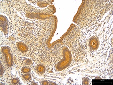
Secreted phosphoprotein 1 (SPP1) is an extracellular matrix glycoprotein that is highly expressed at the maternal–fetal interface and is a critical mediator of embryo implantation. The objectives of this study were to examine the spatial and temporal cyclical expression patterns and steroid regulation of SPP1 mRNA and protein in ovine endometrium, which may be further indicative of their functionality in embryo implantation. Uterine tissue was obtained following hysterectomy from ovariectomised ewes treated with ovarian steroids. In parallel, in vitro culture of endometrial cells was used to investigate the effects of ovarian steroids on SPP1 expression in endometrial and luminal epithelial (LE) cells. A significant sustained mid-luteal phase increase in SPP1 mRNA in intercaruncular regions of the endometrium was observed, indicating that glandular epithelium is likely to be the primary source of SPP1 production. This increase in SPP1 was induced by progesterone treatment and was shown at the protein level by immunohistochemistry analysis. Similarly, treatment of stromal cells with 10 ng mL−1 progesterone or in combination with 1 ng mL−1 oestradiol significantly increased SPP1 expression (P < 0.05). Collectively, expression levels of SPP1 are cycle-dependent and peak in the progesterone-dominant luteal phase. They are dependent on the interaction of uterine LE and stromal cells and may involve paracrine signalling by progesterone receptor-positive stromal cells.
Keywords: OVX, sheep, SPP1, steroid hormones, uterus.
References
Aplin, J. D. (2010). Developmental cell biology of human villous trophoblast: current research problems. Int. J. Dev. Biol. 54, 323–329.| Developmental cell biology of human villous trophoblast: current research problems.Crossref | GoogleScholarGoogle Scholar | 19876840PubMed |
Apparao, K. B., Murray, M. J., Fritz, M. A., Meyer, W. R., Chambers, A. F., Truong, P. R., and Lessey, B. A. (2001). Osteopontin and its receptor alphavbeta(3) integrin are coexpressed in the human endometrium during the menstrual cycle but regulated differentially. J. Clin. Endocrinol. Metab. 86, 4991–5000.
| 11600576PubMed |
Bailey, D. W., Dunlap, K. A., Erikson, D. W., Patel, A. K., Bazer, F. W., Burghardt, R. C., and Johnson, G. A. (2010). Effects of long-term progesterone exposure on porcine uterine gene expression: progesterone alone does not induce secreted phosphoprotein 1 (osteopontin) in glandular epithelium. Reproduction 140, 595–604.
| Effects of long-term progesterone exposure on porcine uterine gene expression: progesterone alone does not induce secreted phosphoprotein 1 (osteopontin) in glandular epithelium.Crossref | GoogleScholarGoogle Scholar | 20705772PubMed |
Berneau, S. C., Ruane, P. T., Brison, D. R., Kimber, S. J., Westwood, M., and Aplin, J. D. (2019). Characterisation of osteopontin in an in vitro model of embryo implantation. Cells 8, 432.
| Characterisation of osteopontin in an in vitro model of embryo implantation.Crossref | GoogleScholarGoogle Scholar |
Casals, G., Ordi, J., Creus, M., Fabregues, F., Carmona, F., Casamitjana, R., and Balasch, J. (2010). Osteopontin and alphavbeta3 integrin as markers of endometrial receptivity: the effect of different hormone therapies. Reprod. Biomed. Online 21, 349–359.
| Osteopontin and alphavbeta3 integrin as markers of endometrial receptivity: the effect of different hormone therapies.Crossref | GoogleScholarGoogle Scholar | 20638909PubMed |
Chaen, T., Konno, T., Egashira, M., Bai, R., Nomura, N., Nomura, S., Hirota, Y., Sakurai, T., and Imakawa, K. (2012). Estrogen-dependent uterine secretion of osteopontin activates blastocyst adhesion competence. PLoS One 7, e48933.
| Estrogen-dependent uterine secretion of osteopontin activates blastocyst adhesion competence.Crossref | GoogleScholarGoogle Scholar | 23152823PubMed |
Dunlap, K. A., Erikson, D. W., Burghardt, R. C., White, F. J., Reed, K. M., Farmer, J. L., Spencer, T. E., Magness, R. R., Bazer, F. W., Bayless, K. J., and Johnson, G. A. (2008). Progesterone and placentation increase secreted phosphoprotein one (SPP1 or osteopontin) in uterine glands and stroma for histotrophic and hematotrophic support of ovine pregnancy. Biol. Reprod. 79, 983–990.
| Progesterone and placentation increase secreted phosphoprotein one (SPP1 or osteopontin) in uterine glands and stroma for histotrophic and hematotrophic support of ovine pregnancy.Crossref | GoogleScholarGoogle Scholar | 18667748PubMed |
DuQuesnay, R., Wright, C., Aziz, A. A., Stamp, G. W., Trew, G. H., Margara, R. A., and White, J. O. (2009). Infertile women with isolated polycystic ovaries are deficient in endometrial expression of osteopontin but not alphavbeta3 integrin during the implantation window. Fertil. Steril. 91, 489–499.
| Infertile women with isolated polycystic ovaries are deficient in endometrial expression of osteopontin but not alphavbeta3 integrin during the implantation window.Crossref | GoogleScholarGoogle Scholar | 18353318PubMed |
Erikson, D. W., Burghardt, R. C., Bayless, K. J., and Johnson, G. A. (2009). Secreted phosphoprotein 1 (SPP1, osteopontin) binds to integrin alpha v beta 6 on porcine trophectoderm cells and integrin alpha v beta 3 on uterine luminal epithelial cells, and promotes trophectoderm cell adhesion and migration. Biol. Reprod. 81, 814–825.
| Secreted phosphoprotein 1 (SPP1, osteopontin) binds to integrin alpha v beta 6 on porcine trophectoderm cells and integrin alpha v beta 3 on uterine luminal epithelial cells, and promotes trophectoderm cell adhesion and migration.Crossref | GoogleScholarGoogle Scholar | 19571258PubMed |
Gray, C. A., Taylor, K. M., Ramsey, W. S., Hill, J. R., Bazer, F. W., Bartol, F. F., and Spencer, T. E. (2001). Endometrial glands are required for preimplantation conceptus elongation and survival. Biol. Reprod. 64, 1608–1613.
| Endometrial glands are required for preimplantation conceptus elongation and survival.Crossref | GoogleScholarGoogle Scholar | 11369585PubMed |
Groll, J. M., Usadi, R. S., Lessey, B. A., Lininger, R., Young, S. L., and Fritz, M. A. (2009). Effects of variations in serum estradiol concentrations on secretory endometrial development and function in experimentally induced cycles in normal women. Fertil. Steril. 92, 2058–2061.
| Effects of variations in serum estradiol concentrations on secretory endometrial development and function in experimentally induced cycles in normal women.Crossref | GoogleScholarGoogle Scholar | 19608171PubMed |
Jackson, G. L., and Kuehl, D. (2004). Effects of applying gamma-aminobutyric acid(B) drugs into the medial basal hypothalamus on basal luteinizing hormone concentrations and on luteinizing hormone surges in the female sheep. Biol. Reprod. 70, 334–339.
| Effects of applying gamma-aminobutyric acid(B) drugs into the medial basal hypothalamus on basal luteinizing hormone concentrations and on luteinizing hormone surges in the female sheep.Crossref | GoogleScholarGoogle Scholar | 14561650PubMed |
Johnson, G. A., Burghardt, R. C., Spencer, T. E., Newton, G. R., Ott, T. L., and Bazer, F. W. (1999). Ovine osteopontin: II. Osteopontin and alpha(v)beta(3) integrin expression in the uterus and conceptus during the periimplantation period. Biol. Reprod. 61, 892–899.
| Ovine osteopontin: II. Osteopontin and alpha(v)beta(3) integrin expression in the uterus and conceptus during the periimplantation period.Crossref | GoogleScholarGoogle Scholar | 10491621PubMed |
Johnson, G. A., Spencer, T. E., Burghardt, R. C., Taylor, K. M., Gray, C. A., and Bazer, F. W. (2000). Progesterone modulation of osteopontin gene expression in the ovine uterus. Biol. Reprod. 62, 1315–1321.
| Progesterone modulation of osteopontin gene expression in the ovine uterus.Crossref | GoogleScholarGoogle Scholar | 10775182PubMed |
Johnson, G. A., Burghardt, R. C., Joyce, M. M., Spencer, T. E., Bazer, F. W., Gray, C. A., and Pfarrer, C. (2003). Osteopontin is synthesized by uterine glands and a 45-kDa cleavage fragment is localized at the uterine–placental interface throughout ovine pregnancy. Biol. Reprod. 69, 92–98.
| Osteopontin is synthesized by uterine glands and a 45-kDa cleavage fragment is localized at the uterine–placental interface throughout ovine pregnancy.Crossref | GoogleScholarGoogle Scholar | 12606367PubMed |
Kang, Y. J., Forbes, K., Carver, J., and Aplin, J. D. (2014). The role of the osteopontin–integrin alphavbeta3 interaction at implantation: functional analysis using three different in vitro models. Hum. Reprod. 29, 739–749.
| The role of the osteopontin–integrin alphavbeta3 interaction at implantation: functional analysis using three different in vitro models.Crossref | GoogleScholarGoogle Scholar | 24442579PubMed |
Kimmins, S., Lim, H. C., and MacLaren, L. A. (2004). Immunohistochemical localization of integrin alpha V beta 3 and osteopontin suggests that they do not interact during embryo implantation in ruminants. Reprod. Biol. Endocrinol. 2, 19.
| Immunohistochemical localization of integrin alpha V beta 3 and osteopontin suggests that they do not interact during embryo implantation in ruminants.Crossref | GoogleScholarGoogle Scholar | 15115551PubMed |
Marei, W. F. A., Wathes, D. C., Raheem, K. A., Mohey-Elsaeed, O., Ghafari, F., and Fouladi-Nashta, A. A. (2017). Influence of hyaluronan on endometrial receptivity and embryo attachment in sheep. Reprod. Fertil. Dev. 29, 1763–1773.
| Influence of hyaluronan on endometrial receptivity and embryo attachment in sheep.Crossref | GoogleScholarGoogle Scholar |
Omigbodun, A., Ziolkiewicz, P., Tessler, C., Hoyer, J. R., and Coutifaris, C. (1997). Progesterone regulates osteopontin expression in human trophoblasts: a model of paracrine control in the placenta? Endocrinology 138, 4308–4315.
| Progesterone regulates osteopontin expression in human trophoblasts: a model of paracrine control in the placenta?Crossref | GoogleScholarGoogle Scholar | 9322944PubMed |
Pandey, S., Ahmad Bhat, I., Kumar Bharti, M., Shabir, U., Ahmad Peer, B., Baiju, I., Sonwane, A., Chandra, V., Sai Kumar, G., and Taru Sharma, G. (2020). Progesterone modulates adhesion molecules in uterine epithelial cells and in vitro embryo production in buffalo. Reprod. Domest. Anim. 55, 833–843.
| Progesterone modulates adhesion molecules in uterine epithelial cells and in vitro embryo production in buffalo.Crossref | GoogleScholarGoogle Scholar | 32335951PubMed |
Peyghambari, F., Salehnia, M., Forouzandeh Moghadam, M., Rezazadeh Valujerdi, M., and Hajizadeh, E. (2010). The correlation between the endometrial integrins and osteopontin expression with pinopodes development in ovariectomized mice in response to exogenous steroids hormones. Iran. Biomed. J. 14, 109–119.
| 21079662PubMed |
Ponglowhapan, S., Church, D. B., and Khalid, M. (2008). Differences in the proportion of collagen and muscle in the canine lower urinary tract with regard to gonadal status and gender. Theriogenology 70, 1516–1524.
| Differences in the proportion of collagen and muscle in the canine lower urinary tract with regard to gonadal status and gender.Crossref | GoogleScholarGoogle Scholar | 18703223PubMed |
Quirke, J. F., Hanrahan, J. P., and Gosling, J. P. (1979). Plasma progesterone levels throughout the oestrous cycle and release of LH at oestrus in sheep with different ovulation rates. J. Reprod. Fertil. 55, 37–44.
| Plasma progesterone levels throughout the oestrous cycle and release of LH at oestrus in sheep with different ovulation rates.Crossref | GoogleScholarGoogle Scholar | 570603PubMed |
Raheem, K. A., Marei, W. F. A., Campbell, B. K., and Fouladi-Nashta, A. A. (2016). In vivo and in vitro studies of MUC1 regulation in sheep endometrium. Theriogenology 85, 1635–1643.
| In vivo and in vitro studies of MUC1 regulation in sheep endometrium.Crossref | GoogleScholarGoogle Scholar | 26934922PubMed |
Robinson, R. S., Pushpakumara, P. G., Cheng, Z., Peters, A. R., Abayasekara, D. R., and Wathes, D. C. (2002). Effects of dietary polyunsaturated fatty acids on ovarian and uterine function in lactating dairy cows. Reproduction 124, 119–131.
| 12090925PubMed |
Satterfield, M. C., Song, G., Hayashi, K., Bazer, F. W., and Spencer, T. E. (2008). Progesterone regulation of the endometrial WNT system in the ovine uterus. Reprod. Fertil. Dev. 20, 935–946.
| Progesterone regulation of the endometrial WNT system in the ovine uterus.Crossref | GoogleScholarGoogle Scholar | 19007558PubMed |
Spencer, T. E., and Bazer, F. W. (2004). Conceptus signals for establishment and maintenance of pregnancy. Reprod. Biol. Endocrinol. 2, 49.
| Conceptus signals for establishment and maintenance of pregnancy.Crossref | GoogleScholarGoogle Scholar | 15236653PubMed |
Spencer, T. E., Becker, W. C., George, P., Mirando, M. A., Ogle, T. F., and Bazer, F. W. (1995). Ovine interferon-tau regulates expression of endometrial receptors for estrogen and oxytocin but not progesterone. Biol. Reprod. 53, 732–745.
| Ovine interferon-tau regulates expression of endometrial receptors for estrogen and oxytocin but not progesterone.Crossref | GoogleScholarGoogle Scholar | 7578700PubMed |
Swangchan-Uthai, T., Lavender, C. R., Cheng, Z., Fouladi-Nashta, A. A., and Wathes, D. C. (2012). Time course of defense mechanisms in bovine endometrium in response to lipopolysaccharide. Biol. Reprod. 87, 135.
| Time course of defense mechanisms in bovine endometrium in response to lipopolysaccharide.Crossref | GoogleScholarGoogle Scholar | 23077171PubMed |
Teklenburg, G., Salker, M., Molokhia, M., Lavery, S., Trew, G., Aojanepong, T., Mardon, H. J., Lokugamage, A. U., Rai, R., Landles, C., Roelen, B. A., Quenby, S., Kuijk, E. W., Kavelaars, A., Heijnen, C. J., Regan, L., Brosens, J. J., and Macklon, N. S. (2010). Natural selection of human embryos: decidualizing endometrial stromal cells serve as sensors of embryo quality upon implantation. PLoS One 5, e10258.
| Natural selection of human embryos: decidualizing endometrial stromal cells serve as sensors of embryo quality upon implantation.Crossref | GoogleScholarGoogle Scholar | 20422017PubMed |
Wang, X. B., Qi, Q. R., Wu, K. L., and Xie, Q. Z. (2018). Role of osteopontin in decidualization and pregnancy success. Reproduction 155, 423–432.
| Role of osteopontin in decidualization and pregnancy success.Crossref | GoogleScholarGoogle Scholar | 29420252PubMed |
Wathes, D. C., and Hamon, M. (1993). Localization of oestradiol, progesterone and oxytocin receptors in the uterus during the oestrous cycle and early pregnancy of the ewe. J. Endocrinol. 138, 479–492.
| Localization of oestradiol, progesterone and oxytocin receptors in the uterus during the oestrous cycle and early pregnancy of the ewe.Crossref | GoogleScholarGoogle Scholar | 8277221PubMed |