

208 The effect of copper on the differentiation of adipose-derived stem cells into osteoblasts
T. Bane A , L. Siegel A , J. Bertels A , K. Ratz A , M. Rubessa A and M. Wheeler A BA Department of Animal Science University of Illinois, Urbana, IL, USA;
B Carl R. Woese Institute of Genomic Biology University of Illinois, Urbana, IL, USA
Reproduction, Fertility and Development 31(1) 229-230 https://doi.org/10.1071/RDv31n1Ab208
Published online: 3 December 2018
Abstract
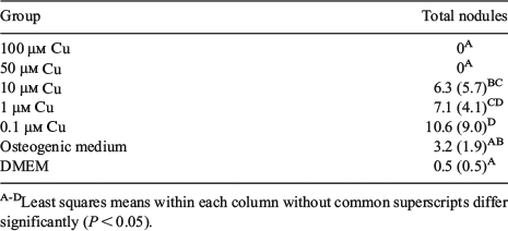
Large bone defects present a tremendous challenge to the treating surgeon. Tissue engineering using scaffolds of various sizes and shapes that contain stem cells and other osteoinductive molecules offer a potential solution to this difficult problem. The aim of this project was to evaluate whether osteogenic medium infused with copper influences the differentiation of adipose-derived stem cells (ASC) into osteoblasts. Copper is a key cofactor for lysyl oxidase, an enzyme involved in producing a collagen matrix through which bone can grow. Lysyl oxidase expression is up-regulated in bone marrow stromal cells (Khosravi et al. 2014 PLoS One 9, e100669). Our hypothesis was that the presence of copper in the osteogenic medium would positively influence the number of osteoblastic nodules formed. Swine ASC were isolated as described (Monaco et al. 2009 Open Tissue Eng. Regen. Med. J. 2, 20-33). The ASC were divided in 7 different treatments: 5 concentrations of copper in the osteogenic medium (0.1, 1, 10, 50, and 100 µM) plus 2 control treatments (osteogenic medium without copper and a negative control, DMEM). The medium was changed twice a week for 4 weeks. The experiment was replicated 6 times. At the end of the culture period, cells were stained with Alizarin Red S and Von Kossa stains. In each well, we counted the total number of nodules that were either formed or forming. Data were analysed using the generalized linear model (GLM) procedure (SPSS Inc./IBM Corp., Armonk, NY). The least significant difference (l.s.d.) post hoc test was used to perform statistical multiple comparison. The α-level was set at 0.05. The results showed that more nodules were formed in the 0.1 and 1 µM copper groups compared with the osteogenic control, but there was no statistical difference between those 2 treatments. Table 1 illustrates the total number of formed and forming nodules in addition to their standard deviation. There was a positive effect on nodule formation when copper concentrations of 0.1 and 1 µM were added to the osteogenic medium. In contrast, copper concentrations of 50 and 100 µM had a cytotoxic effect. These results confirm that low concentrations of copper have a positive effect on osteogenesis. This preliminary experiment is the first step towards the analysis of the behaviour of ASC on scaffolds with copper incorporated into their matrix.
|