

24 EFFECT OF TREATMENT WITH TRICHOSTATIN A ON IN VITRO DEVELOPMENT OF BOVINE NUCLEAR-TRANSFERRED EMBRYOS
S. Akagi A , K. Fukunari B , K. Matsukawa A , S. Watanabe A and S. Takahashi AA National Institute of Livestock and Grassland Science, Tsukuba, Ibaraki, Japan
B Iwate Agricultural Research Center, Takizawa, Iwate, Japan
Reproduction, Fertility and Development 19(1) 130-131 https://doi.org/10.1071/RDv19n1Ab24
Submitted: 12 October 2006 Accepted: 12 October 2006 Published: 12 December 2006
Abstract
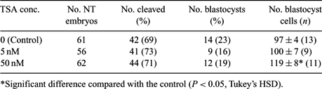
It has been reported that 5 or 50 nM trichostatin A (TSA) treatment after somatic cell nuclear transfer (NT) improves the success rate of mouse cloning (Kishigami et al. 2006 Biochem. Biophys. Res. Commun. 340, 183–189). In this study, we examined the effect of TSA treatment on the in vitro development of bovine NT embryos. As donor cells for NT, bovine fibroblast cells of passages 3 to 5 were used following culture in serum-starved medium for 5 to 7 days. Oocytes were enucleated after in vitro maturation in TCM-199 supplemented with 10% fetal bovine serum. Enucleated MII oocytes were fused with fibroblast cells by a DC pulse of 25 V/150 µm for 10 µs in Zimmerman mammalian cell fusion medium. Fused oocytes were activated by 10 µM calcium ionophore for 5 min, followed by incubation with 2.5 µg mL−1 cytochalasin D, 10 µg mL−1 cycloheximide, and 5 or 50 nM TSA for 1 h, and then cycloheximide and 5 or 50 nM TSA for 4 h. After chemical activation, NT embryos were cultured in IVD-101 (Research Institute of Functional Peptide Co., Ltd., Yamagata, Japan) with 5 or 50 nM TSA for 10 h and subsequently cultured in IVD-101 without TSA. Control NT embryos were cultured in the same medium without TSA after fusion. After in vitro culture for 8 days, blastocyst formation and cell numbers of blastocysts were examined. The fusion rate of enucleated oocytes with fibroblast cells was 81% (199/247). In vitro development of NT embryos is summarized in Table 1. There were no differences in the cleavage rate and development rate to the blastocyst stage of NT embryos among control, and 5 and 50 nM TSA treatments. The cell number of 50 nM TSA-treated NT embryos at the blastocyst stage was higher than that of control NT embryos without TSA treatment. In conclusion, 50 nM TSA treatment for 15 h after activation did not affect the in vitro developmental competence, but increased total cell number in bovine NT embryos. These results suggest that TSA treatment may improve the quality of blastocysts in bovine NT.
|