Elucidation of the complex carbohydrate structures of canola meal fibre by commercial feed enzymes
N. R. Pedersen A , K. Stamatopoulos B C , D. Pettersson A , K. A. Kristensen A and J. L. Ravn AA Animal Health and Nutrition, Novozymes A/S, 2880 Denmark.
B DSM Nutritional Products Australia Pty Ltd, Wagga Wagga, NSW 2650.
C Corresponding author. Email: Kostas.stamatopoulos@dsm.com
Animal Production Science 57(12) 2435-2435 https://doi.org/10.1071/ANv57n12Ab137
Published: 20 November 2017
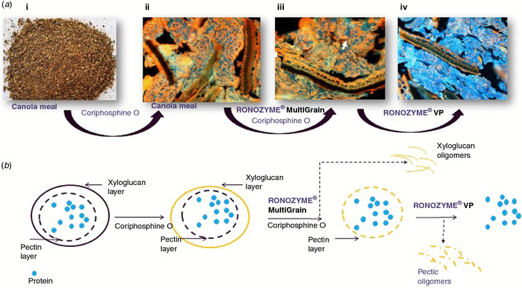
Inclusion levels of canola meal (CM) in pig diets may be restricted by the concentration of glucosinolates (Schone et al. 2001) and by relatively high concentration of fibre (plant cell walls) in the meal. However, new varieties of canola containing more protein and less fibre than conventional CM have been identified. Due to the likely higher total glucosinolate levels in the high protein CM (CM-HP) variety (15 μmol/g) compared to the conventional CM (CM-CV) variety (8.69 μmol/g), it was observed that the standardised ileal digestibility of amino acids in pigs fed with CM-HP from black-seeded canola was not greater than in CM-CV (Berrocoso et al. 2015). The high fibre components in CM include mainly non-cellulosic polysaccharides (13–16%). The fibre in CM is antinutritional and is responsible for the less than optimum digestibility values for constituents such as protein (70–75%) (Sauer et al. 1980). Since non-ruminants do not possess fibre degrading enzymes, use of exogenous enzymes seems to be essential. In vitro microscopy work elucidating the cell wall structure of soybean and the effect of enzymes on the same has been published earlier (Ravn et al. 2015). In the current in vitro work, using techniques devised by Ravn et al. (2015), the complex fibre structure of canola/CM and the impact of enzymes was examined. Using a dye staining acidic polysaccharides orange (Coriphosphine O, TCI America, Portland, OR, USA) and antibodies targeting plant cell wall structures, identification of the cell wall of canola was possible, showing that the structure of cell wall of canola is different from that of soybean (Ravn et al. 2015). Using a cell wall specific antibody recognising xyloglucan epitopes, a pure commercial xyloglucanase from Megazyme International Ltd (Co. Wicklow, Ireland) and a commercial feed xyloglucanase (Ronozyme MultiGrain, DSM, Wagga Wagga, NSW, Australia), the outmost cell wall layer of canola was identified as xyloglucan (Fig. 1Aii). Removal of the xyloglucan layer after incubation at room temperature for 3 h by either enzyme, washing the sample and re-staining with either Coriphosphine O (Fig. 1Aiii) or use of antibodies revealed a pectin layer below the xyloglucan layer. (Ronozyme VP, DSM, Wagga Wagga, NSW, Australia) (containing pectinase) removal of the pectin layer revealed protein underneath, indicating protein accessibility on removal of cell wall fibre (Fig. 1Aiv). Each experiment was repeated three times.
This research on elucidation of cell wall morphology of differing protein sources and the use of enzymes to degrade the same can be used to highlight the importance of using the right combination of enzymes. In turn this may assist in increasing nutritional worth of canola and reduce feed costs while maintaining performance.
References
Berrocoso JD, Rojas OJ, Liu Y, Shoulders J, Gonzalez-Vega JC, Stein HH (2015) Journal of Animal Science 93, 2208–2217.| Crossref | GoogleScholarGoogle Scholar |
Ravn JL, Martens HJ, Pettersson D, Pedersen NR (2015) Journal of Agricultural Science 7, 1–13.
Sauer WC, Just A, Jørgensen H, Fekadu M, Eggum BO (1980) Acta Agriculturae Scandinavica 30, 449–459.
| Crossref | GoogleScholarGoogle Scholar |
Schöne F, Leiterer M, Hartung H, Jahreis G, Tischendorf F (2001) British Journal of Nutrition 85, 659–670.
| Crossref | GoogleScholarGoogle Scholar |


